 Movie
Movie Controller

[English] 日本語
 Yorodumi
- PDB-1hsr: BINDING MODE OF BENZHYDROXAMIC ACID TO ARTHROMYCES RAMOSUS PEROXIDASE -
+ Open data

- Basic information
| Entry | Database: PDB / ID: 1hsr | |||||||||
|---|---|---|---|---|---|---|---|---|---|---|
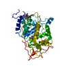
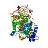
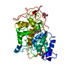
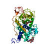
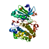
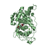
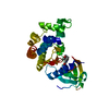
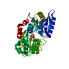
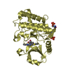
| Title | BINDING MODE OF BENZHYDROXAMIC ACID TO ARTHROMYCES RAMOSUS PEROXIDASE | |||||||||
 Components Components | PEROXIDASE | |||||||||
 Keywords Keywords | OXIDOREDUCTASE / GLYCOPROTEIN / PEROXIDASE | |||||||||
| Function / homology |  Function and homology information Function and homology informationlactoperoxidase activity / peroxidase / hydrogen peroxide catabolic process / response to reactive oxygen species / cellular response to oxidative stress / heme binding / extracellular region / metal ion binding Similarity search - Function | |||||||||
| Biological species |  'Arthromyces ramosus' (fungus) 'Arthromyces ramosus' (fungus) | |||||||||
| Method |  X-RAY DIFFRACTION / SYNCHROTRON / Resolution: 1.6 Å X-RAY DIFFRACTION / SYNCHROTRON / Resolution: 1.6 Å | |||||||||
 Authors Authors | Fukuyama, K. / Itakura, H. | |||||||||
 Citation Citation | Journal: FEBS Lett. / Year: 1997 Title: Binding mode of benzhydroxamic acid to Arthromyces ramosus peroxidase shown by X-ray crystallographic analysis of the complex at 1.6 A resolution. Authors: Itakura, H. / Oda, Y. / Fukuyama, K. #1: Journal: J.Biol.Chem. / Year: 1997Title: Binding of Iodide to Arthromyces Ramosus Peroxidase Investigated with X-Ray Crystallographic Analysis, 1H and 127I NMR Spectroscopy, and Steady-State Kinetics Authors: Fukuyama, K. / Sato, K. / Itakura, H. / Takahashi, S. / Hosoya, T. #2: Journal: FEBS Lett. / Year: 1996Title: Pentacoordination of the Heme Iron of Arthromyces Ramosus Peroxidase Shown by a 1.8 A Resolution Crystallographic Study at Ph 4.5 Authors: Kunishima, N. / Amada, F. / Fukuyama, K. / Kawamoto, M. / Matsunaga, T. / Matsubara, H. #3: Journal: J.Biol.Chem. / Year: 1995Title: Crystal Structures of Cyanide-and Triiodide-Bound Forms of Arthromyces Ramosus Peroxidase at Different Ph Values. Perturbations of Active Site Residues and Their Implication in Enzyme Catalysis Authors: Fukuyama, K. / Kunishima, N. / Amada, F. / Kubota, T. / Matsubara, H. #4: Journal: J.Mol.Biol. / Year: 1994Title: Crystal Structure of the Fungal Peroxidase from Arthromyces Ramosus at 1.9 A Resolution. Structural Comparisons with the Lignin and Cytochrome C Peroxidases Authors: Kunishima, N. / Fukuyama, K. / Matsubara, H. / Hatanaka, H. / Shibano, Y. / Amachi, T. #5: Journal: Proteins / Year: 1993Title: Crystallization and Preliminary X-Ray Diffraction Studies of Peroxidase from a Fungus Arthromyces Ramosus Authors: Kunishima, N. / Fukuyama, K. / Wakabayashi, S. / Sumida, M. / Takaya, M. / Shibano, Y. / Amachi, T. / Matsubara, H. | |||||||||
| History |
|
- Structure visualization
| Structure viewer | Molecule: MolmilJmol/JSmol |
|---|
- Downloads & links
-Download
| PDBx/mmCIF format | 1hsr.cif.gz | 84 KB | Display | PDBx/mmCIF format |
|---|---|---|---|---|
| PDB format | pdb1hsr.ent.gz | 61.1 KB | Display | PDB format |
| PDBx/mmJSON format | 1hsr.json.gz | Tree view | PDBx/mmJSON format | |
| Others |  Other downloads Other downloads |
-Validation report
| Arichive directory | https://data.pdbj.org/pub/pdb/validation_reports/hs/1hsrftp://data.pdbj.org/pub/pdb/validation_reports/hs/1hsr | HTTPS FTP |
|---|
-Related structure data
| Similar structure data |
|---|

-Links
 PDBj
PDBj
- Assembly
| Deposited unit | 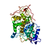
| ||||||||
|---|---|---|---|---|---|---|---|---|---|
| 1 |
| ||||||||
| Unit cell |
|
-Components
-Protein / Sugars , 2 types, 2 molecules A
| #1: Protein | Mass: 35722.797 Da / Num. of mol.: 1 / Source method: isolated from a natural source / Source: (natural) 'Arthromyces ramosus' (fungus) / Genus: 'Arthromyces' / References: UniProt: P28313, peroxidase |
|---|---|
| #2: Polysaccharide | 2-acetamido-2-deoxy-beta-D-glucopyranose-(1-4)-2-acetamido-2-deoxy-beta-D-glucopyranose Source method: isolated from a genetically manipulated source |
-Non-polymers , 4 types, 245 molecules 


| #3: Chemical |  Mass: 40.078 Da / Num. of mol.: 2 / Source method: obtained synthetically / Formula: Ca Mass: 40.078 Da / Num. of mol.: 2 / Source method: obtained synthetically / Formula: Ca#4: Chemical | ChemComp-HEM / |  Mass: 616.487 Da / Num. of mol.: 1 / Source method: obtained synthetically / Formula: C34H32FeN4O4 Mass: 616.487 Da / Num. of mol.: 1 / Source method: obtained synthetically / Formula: C34H32FeN4O4#5: Chemical | ChemComp-BHO / |  Mass: 137.136 Da / Num. of mol.: 1 / Source method: obtained synthetically / Formula: C7H7NO2 Mass: 137.136 Da / Num. of mol.: 1 / Source method: obtained synthetically / Formula: C7H7NO2#6: Water | ChemComp-HOH / | Mass: 18.015 Da / Num. of mol.: 241 / Source method: isolated from a natural source / Formula: H2O |
|---|
-Details
| Has protein modification | Y |
|---|
-Experimental details
-Experiment
| Experiment | Method: X-RAY DIFFRACTION |
|---|
- Sample preparation
| Crystal | Density Matthews: 2.29 Å3/Da / Density % sol: 46 % Description: DATA WERE COLLECTED ON A WEISSENBERG CAMERA USING A CYLINDRICAL CASSETTE HAVING A 287 MM RADIUS. | ||||||||||||||||||||
|---|---|---|---|---|---|---|---|---|---|---|---|---|---|---|---|---|---|---|---|---|---|
| Crystal grow | pH: 5.5 / Details: pH 5.5 | ||||||||||||||||||||
| Crystal | *PLUS | ||||||||||||||||||||
| Crystal grow | *PLUS Temperature: 22-24 ℃ / pH: 7.5 / Method: vapor diffusion, hanging dropDetails: repeated seeding, Kunishima, N., (1993) Proteins: Struct.,Funct., Genet., 15, 216. | ||||||||||||||||||||
| Components of the solutions | *PLUS
|
-Data collection
| Diffraction source | Source: SYNCHROTRON / Site: Photon Factory  / Beamline: BL-6A / Wavelength: 1 / Beamline: BL-6A / Wavelength: 1 |
|---|---|
| Detector | Type: FUJI / Detector: IMAGE PLATE |
| Radiation | Monochromatic (M) / Laue (L): M / Scattering type: x-ray |
| Radiation wavelength | Wavelength: 1 Å / Relative weight: 1 |
| Reflection | *PLUS Highest resolution: 1.5 Å / Num. obs: 47193 / % possible obs: 87.6 % / Num. measured all: 225152 / Rmerge(I) obs: 0.047 |
- Processing
| Software |
| ||||||||||||||||||||||||||||||||||||||||||||||||||||||||||||
|---|---|---|---|---|---|---|---|---|---|---|---|---|---|---|---|---|---|---|---|---|---|---|---|---|---|---|---|---|---|---|---|---|---|---|---|---|---|---|---|---|---|---|---|---|---|---|---|---|---|---|---|---|---|---|---|---|---|---|---|---|---|
| Refinement | Resolution: 1.6→7 Å / Rfactor Rwork: 0.187 / Rfactor obs: 0.187 | ||||||||||||||||||||||||||||||||||||||||||||||||||||||||||||
| Refinement step | Cycle: LAST / Resolution: 1.6→7 Å
| ||||||||||||||||||||||||||||||||||||||||||||||||||||||||||||
| Refine LS restraints |
| ||||||||||||||||||||||||||||||||||||||||||||||||||||||||||||
| Refinement | *PLUS Num. reflection obs: 39924 / σ(F): 2 / Rfactor Rfree: 0.227 | ||||||||||||||||||||||||||||||||||||||||||||||||||||||||||||
| Solvent computation | *PLUS | ||||||||||||||||||||||||||||||||||||||||||||||||||||||||||||
| Displacement parameters | *PLUS | ||||||||||||||||||||||||||||||||||||||||||||||||||||||||||||
| Refine LS restraints | *PLUS
|
