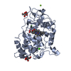
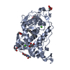
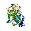
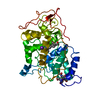
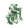
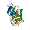
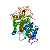
lactoperoxidase activity / peroxidase / hydrogen peroxide catabolic process / response to reactive oxygen species / cellular response to oxidative stress / heme binding / extracellular region / metal ion binding Similarity search - Function
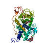
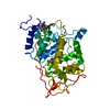
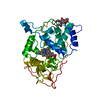
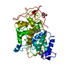
#1: Journal: FEBS Lett. / Year: 1994 Title: Three-Dimensional Structure of a Recombinant Peroxidase from Coprinus Cinereus at 2.6 A Authors: Petersen, J.F.W. / Kadziola, A. / Larsen, S.
History
Deposition
Sep 5, 2002
Deposition site: PDBE / Processing site: PDBE
Revision 1.0
Jun 12, 2003
Provider: repository / Type: Initial release
Revision 1.1
Oct 19, 2011
Group: Derived calculations / Non-polymer description ...Derived calculations / Non-polymer description / Other / Version format compliance
Revision 1.2
May 22, 2019
Group: Data collection / Other / Refinement description Category: pdbx_database_proc / pdbx_database_status / refine Item: _pdbx_database_status.recvd_author_approval / _refine.pdbx_ls_cross_valid_method
In the structure databanks used in Yorodumi, some data are registered as the other names, "COVID-19 virus" and "2019-nCoV". Here are the details of the virus and the list of structure data.
Jan 31, 2019. EMDB accession codes are about to change! (news from PDBe EMDB page)
EMDB accession codes are about to change! (news from PDBe EMDB page)
The allocation of 4 digits for EMDB accession codes will soon come to an end. Whilst these codes will remain in use, new EMDB accession codes will include an additional digit and will expand incrementally as the available range of codes is exhausted. The current 4-digit format prefixed with “EMD-” (i.e. EMD-XXXX) will advance to a 5-digit format (i.e. EMD-XXXXX), and so on. It is currently estimated that the 4-digit codes will be depleted around Spring 2019, at which point the 5-digit format will come into force.
The EM Navigator/Yorodumi systems omit the EMD- prefix.
Related info.:Q: What is EMD? / ID/Accession-code notation in Yorodumi/EM Navigator
Yorodumi is a browser for structure data from EMDB, PDB, SASBDB, etc.
This page is also the successor to EM Navigator detail page, and also detail information page/front-end page for Omokage search.
The word "yorodu" (or yorozu) is an old Japanese word meaning "ten thousand". "mi" (miru) is to see.
Related info.:EMDB / PDB / SASBDB / Comparison of 3 databanks / Yorodumi Search / Aug 31, 2016. New EM Navigator & Yorodumi / Yorodumi Papers / Jmol/JSmol / Function and homology information / Changes in new EM Navigator and Yorodumi
 Movie
Movie Controller

 Yorodumi
 Open data
 Basic information
 Components
 Keywords
 Function and homology information
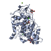
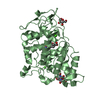
 Coprinopsis cinerea (fungus)
 X-RAY DIFFRACTION /
  Authors
 Citation
 Structure visualization
 Downloads & links
 Other downloads


 PDBj
PDBj
 Assembly
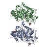

 Type: D-saccharide, beta linking / Mass: 180.156 Da / Num. of mol.: 2


 Mass: 616.487 Da / Num. of mol.: 2 / Source method: obtained synthetically / Formula: C34H32FeN4O4
 Mass: 40.078 Da / Num. of mol.: 4 / Source method: obtained synthetically / Formula: Ca
 Mass: 24.305 Da / Num. of mol.: 1 / Source method: obtained synthetically / Formula: Mg
 Sample preparation
 Processing