 Movie
Movie Controller

[English] 日本語
 Yorodumi
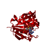
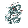
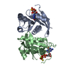
- PDB-2e2a: ASP81LEU ENZYME IIA FROM THE LACTOSE SPECIFIC PTS FROM LACTOCOCCU... -
+ Open data

- Basic information
| Entry | Database: PDB / ID: 2e2a | ||||||
|---|---|---|---|---|---|---|---|
| Title | ASP81LEU ENZYME IIA FROM THE LACTOSE SPECIFIC PTS FROM LACTOCOCCUS LACTIS | ||||||
 Components Components | PROTEIN (ENZYME IIA) | ||||||
 Keywords Keywords | TRANSFERASE / ENZYME IIA / HELICAL BUNDLES / PTS / PHOSPHOTRANSFERASE SYSTEM | ||||||
| Function / homology |  Function and homology information Function and homology informationphosphoenolpyruvate-dependent sugar phosphotransferase system / transferase activity / metal ion binding / cytoplasm Similarity search - Function | ||||||
| Biological species |  Lactococcus lactis (lactic acid bacteria) Lactococcus lactis (lactic acid bacteria) | ||||||
| Method |  X-RAY DIFFRACTION / SYNCHROTRON / OTHER / Resolution: 2.1 Å X-RAY DIFFRACTION / SYNCHROTRON / OTHER / Resolution: 2.1 Å | ||||||
 Authors Authors | Sliz, P. / Koch, B. / Hengstenberg, W. / Pai, E.F. | ||||||
 Citation Citation | Journal: To be Published Title: Structure of Asp81Leu Enzyme Iia from the Lactose Specific Pts from Lactococcus Lactis Authors: Sliz, P. / KOCH, B. / Hengstenberg, W. / Pai, E.F. #1: Journal: Structure / Year: 1997Title: The Structure of Enzyme Iialactose from Lactococcus Lactis Reveals a New Fold and Points to Possible Interactions of a Multicomponent System Authors: Sliz, P. / Engelmann, R. / Hengstenberg, W. / Pai, E.F. | ||||||
| History |
|
- Structure visualization
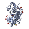
| Structure viewer | Molecule: MolmilJmol/JSmol |
|---|
- Downloads & links
-Download
| PDBx/mmCIF format | 2e2a.cif.gz | 71.7 KB | Display | PDBx/mmCIF format |
|---|---|---|---|---|
| PDB format | pdb2e2a.ent.gz | 54.4 KB | Display | PDB format |
| PDBx/mmJSON format | 2e2a.json.gz | Tree view | PDBx/mmJSON format | |
| Others |  Other downloads Other downloads |
-Validation report
| Arichive directory | https://data.pdbj.org/pub/pdb/validation_reports/e2/2e2aftp://data.pdbj.org/pub/pdb/validation_reports/e2/2e2a | HTTPS FTP |
|---|
-Related structure data
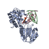
| Related structure data | 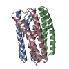 1e2aS S: Starting model for refinement |
|---|---|
| Similar structure data |


-Links
 PDBj
PDBj- Assembly
| Deposited unit | 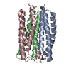
| ||||||||||||||||
|---|---|---|---|---|---|---|---|---|---|---|---|---|---|---|---|---|---|
| 1 |
| ||||||||||||||||
| Unit cell |
| ||||||||||||||||
| Noncrystallographic symmetry (NCS) | NCS oper:
|
-Components
| #1: Protein | Mass: 11461.188 Da / Num. of mol.: 3 / Mutation: D81L Source method: isolated from a genetically manipulated source Source: (gene. exp.) Lactococcus lactis (lactic acid bacteria)Strain: MG1820 / Description: NIZO, NETHERLANDS / Gene: LACF / Production host: References: UniProt: P23532, protein-Npi-phosphohistidine-sugar phosphotransferase #2: Water | ChemComp-HOH / |  Mass: 18.015 Da / Num. of mol.: 173 / Source method: isolated from a natural source / Formula: H2O Mass: 18.015 Da / Num. of mol.: 173 / Source method: isolated from a natural source / Formula: H2O |
|---|
-Experimental details
-Experiment
| Experiment | Method: X-RAY DIFFRACTION / Number of used crystals: 1 |
|---|
- Sample preparation
| Crystal | Density Matthews: 2.25 Å3/Da / Density % sol: 44.56 % |
|---|---|
| Crystal grow | pH: 6.5 Details: THE PROTEIN WAS CRYSTALLIZED FROM 0.9M NA ACETATE AND 0.1M NA CACODYLATE AT PH 6.5. HANGING-DROP METHOD WAS USED. THE INITIAL CONCENTRATION OF THE PROTEIN IN THE DROP WAS 5 MG/ML. |
-Data collection
| Diffraction | Mean temperature: 100 K |
|---|---|
| Diffraction source | Source: SYNCHROTRON / Site: NSLS  / Beamline: X12B / Wavelength: 0.949 / Beamline: X12B / Wavelength: 0.949 |
| Detector | Type: ADSC QUANTUM 4 / Detector: CCD / Date: Mar 30, 1998 |
| Radiation | Protocol: SINGLE WAVELENGTH / Monochromatic (M) / Laue (L): M / Scattering type: x-ray |
| Radiation wavelength | Wavelength: 0.949 Å / Relative weight: 1 |
| Reflection | Resolution: 2.1→36 Å / Num. obs: 18726 / % possible obs: 99.5 % / Observed criterion σ(I): 0 / Redundancy: 4.8 % / Biso Wilson estimate: 30.6 Å2 / Rsym value: 0.037 / Net I/σ(I): 13.7 |
| Reflection shell | Resolution: 2.1→2.2 Å / Redundancy: 4.2 % / Mean I/σ(I) obs: 2.7 / Rsym value: 0.271 / % possible all: 99.9 |
- Processing
| Software |
| ||||||||||||||||||||||||||||||||||||||||||||||||||||||||||||||||||||||||||||||||
|---|---|---|---|---|---|---|---|---|---|---|---|---|---|---|---|---|---|---|---|---|---|---|---|---|---|---|---|---|---|---|---|---|---|---|---|---|---|---|---|---|---|---|---|---|---|---|---|---|---|---|---|---|---|---|---|---|---|---|---|---|---|---|---|---|---|---|---|---|---|---|---|---|---|---|---|---|---|---|---|---|---|
| Refinement | Method to determine structure: OTHER Starting model: 1E2A Resolution: 2.1→20 Å / Rfactor Rfree error: 0.006 / Data cutoff high rms absF: 1666967.76 / Isotropic thermal model: RESTRAINED / Cross valid method: THROUGHOUT / σ(F): 0
| ||||||||||||||||||||||||||||||||||||||||||||||||||||||||||||||||||||||||||||||||
| Solvent computation | Solvent model: FLAT MODEL / Bsol: 59.47 Å2 / ksol: 0.3684 e/Å3 | ||||||||||||||||||||||||||||||||||||||||||||||||||||||||||||||||||||||||||||||||
| Displacement parameters | Biso mean: 45.9 Å2
| ||||||||||||||||||||||||||||||||||||||||||||||||||||||||||||||||||||||||||||||||
| Refine analyze |
| ||||||||||||||||||||||||||||||||||||||||||||||||||||||||||||||||||||||||||||||||
| Refinement step | Cycle: LAST / Resolution: 2.1→20 Å
| ||||||||||||||||||||||||||||||||||||||||||||||||||||||||||||||||||||||||||||||||
| Refine LS restraints |
| ||||||||||||||||||||||||||||||||||||||||||||||||||||||||||||||||||||||||||||||||
| LS refinement shell | Resolution: 2.1→2.23 Å / Rfactor Rfree error: 0.017 / Total num. of bins used: 6
| ||||||||||||||||||||||||||||||||||||||||||||||||||||||||||||||||||||||||||||||||
| Xplor file |
|
