 Movie
Movie Controller


+ Open data

- Basic information
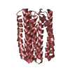
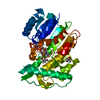
| Entry | Database: PDB / ID: 1e2a | ||||||
|---|---|---|---|---|---|---|---|
| Title | ENZYME IIA FROM THE LACTOSE SPECIFIC PTS FROM LACTOCOCCUS LACTIS | ||||||
 Components Components | ENZYME IIA | ||||||
 Keywords Keywords | TRANSFERASE / ENZYME IIA / HELICAL BUNDLES / PTS / PHOSPHOTRANSFERASE SYSTEM | ||||||
| Function / homology |  Function and homology information Function and homology informationphosphoenolpyruvate-dependent sugar phosphotransferase system / transferase activity / metal ion binding / cytoplasm Similarity search - Function | ||||||
| Biological species |  Lactococcus lactis (lactic acid bacteria) Lactococcus lactis (lactic acid bacteria) | ||||||
| Method |  X-RAY DIFFRACTION / SYNCHROTRON / SINGLE ISOMORPHOUS REPLACEMENT, ANOMALOUS SCATTERING (SIRAS) / Resolution: 2.3 Å X-RAY DIFFRACTION / SYNCHROTRON / SINGLE ISOMORPHOUS REPLACEMENT, ANOMALOUS SCATTERING (SIRAS) / Resolution: 2.3 Å | ||||||
 Authors Authors | Sliz, P. / Engelmann, R. / Hengstenberg, W. / Pai, E.F. | ||||||
 Citation Citation | Journal: Structure / Year: 1997 Title: The structure of enzyme IIAlactose from Lactococcus lactis reveals a new fold and points to possible interactions of a multicomponent system. Authors: Sliz, P. / Engelmann, R. / Hengstenberg, W. / Pai, E.F. #1: Journal: Acta Crystallogr.,Sect.D / Year: 1996Title: Crystallization and Preliminary Structural Studies of Lactose-Specific Enzyme Iia from Lactococcus Lactis Authors: Sliz, P. / Schorter, K.H. / De Vos, W.M. / Pai, E.F. | ||||||
| History |
|
- Structure visualization
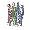
| Structure viewer | Molecule: MolmilJmol/JSmol |
|---|
- Downloads & links
-Download
| PDBx/mmCIF format | 1e2a.cif.gz | 69.7 KB | Display | PDBx/mmCIF format |
|---|---|---|---|---|
| PDB format | pdb1e2a.ent.gz | 52.9 KB | Display | PDB format |
| PDBx/mmJSON format | 1e2a.json.gz | Tree view | PDBx/mmJSON format | |
| Others |  Other downloads Other downloads |
-Validation report
| Arichive directory | https://data.pdbj.org/pub/pdb/validation_reports/e2/1e2aftp://data.pdbj.org/pub/pdb/validation_reports/e2/1e2a | HTTPS FTP |
|---|
-Related structure data
| Similar structure data |
|---|


-Links
 PDBj
PDBj- Assembly
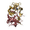
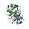
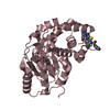
| Deposited unit | 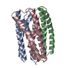
| ||||||||||||||||
|---|---|---|---|---|---|---|---|---|---|---|---|---|---|---|---|---|---|
| 1 |
| ||||||||||||||||
| 2 | 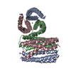
| ||||||||||||||||
| Unit cell |
| ||||||||||||||||
| Noncrystallographic symmetry (NCS) | NCS oper:
|
-Components
| #1: Protein | Mass: 11463.118 Da / Num. of mol.: 3 Source method: isolated from a genetically manipulated source Source: (gene. exp.) Lactococcus lactis (lactic acid bacteria)Strain: MG1820 / Gene: LACF / Production host: References: UniProt: P23532, protein-Npi-phosphohistidine-sugar phosphotransferase #2: Chemical | ChemComp-MG / |   Mass: 24.305 Da / Num. of mol.: 1 / Source method: obtained synthetically / Formula: Mg Mass: 24.305 Da / Num. of mol.: 1 / Source method: obtained synthetically / Formula: Mg#3: Water | ChemComp-HOH / |  Mass: 18.015 Da / Num. of mol.: 56 / Source method: isolated from a natural source / Formula: H2O Mass: 18.015 Da / Num. of mol.: 56 / Source method: isolated from a natural source / Formula: H2O |
|---|
-Experimental details
-Experiment
| Experiment | Method: X-RAY DIFFRACTION / Number of used crystals: 2 |
|---|
- Sample preparation
| Crystal | Density Matthews: 2.48 Å3/Da / Density % sol: 48 % | ||||||||||||||||||||
|---|---|---|---|---|---|---|---|---|---|---|---|---|---|---|---|---|---|---|---|---|---|
| Crystal grow | Method: vapor diffusion, sitting drop / pH: 6.4 Details: CRYSTALS WERE OBTAINED BY THE SITTING-DROP VAPOR-DIFFUSION TECHNIQUE AT ROOM TEMPERATURE USING A SMALL INCREASE IN THE PHOSPHATE BUFFER. THE PROTEIN SOLUTION WAS MIXED WITH 0.15M NA/K ...Details: CRYSTALS WERE OBTAINED BY THE SITTING-DROP VAPOR-DIFFUSION TECHNIQUE AT ROOM TEMPERATURE USING A SMALL INCREASE IN THE PHOSPHATE BUFFER. THE PROTEIN SOLUTION WAS MIXED WITH 0.15M NA/K PHOSPHATE BUFFER, PH 6.4. THE LARGEST CRYSTALS APPEARED IN CIRCA A WEEK., vapor diffusion - sitting drop Temp details: room temp | ||||||||||||||||||||
| Crystal | *PLUS | ||||||||||||||||||||
| Crystal grow | *PLUS Temperature: 25 ℃ / Method: vapor diffusion, sitting drop | ||||||||||||||||||||
| Components of the solutions | *PLUS
|
-Data collection
| Diffraction | Mean temperature: 298 K |
|---|---|
| Diffraction source | Source: SYNCHROTRON / Site: EMBL/DESY, HAMBURG  / Beamline: X31 / Wavelength: 0.94 / Beamline: X31 / Wavelength: 0.94 |
| Detector | Type: MARRESEARCH / Detector: IMAGE PLATE / Date: Oct 1, 1995 |
| Radiation | Monochromatic (M) / Laue (L): M / Scattering type: x-ray |
| Radiation wavelength | Wavelength: 0.94 Å / Relative weight: 1 |
| Reflection | Resolution: 2.3→15 Å / Num. obs: 16632 / % possible obs: 97.7 % / Observed criterion σ(I): 0 / Redundancy: 6.1 % / Rsym value: 0.086 / Net I/σ(I): 18.3 |
| Reflection shell | Resolution: 2.3→2.4 Å / Redundancy: 4.9 % / Mean I/σ(I) obs: 4.9 / Rsym value: 0.36 / % possible all: 98.9 |
| Reflection | *PLUS Rmerge(I) obs: 0.086 |
| Reflection shell | *PLUS % possible obs: 98.9 % / Rmerge(I) obs: 0.363 |
- Processing
| Software |
| ||||||||||||||||||||||||||||||||||||||||||||||||||||||||||||
|---|---|---|---|---|---|---|---|---|---|---|---|---|---|---|---|---|---|---|---|---|---|---|---|---|---|---|---|---|---|---|---|---|---|---|---|---|---|---|---|---|---|---|---|---|---|---|---|---|---|---|---|---|---|---|---|---|---|---|---|---|---|
| Refinement | Method to determine structure: SINGLE ISOMORPHOUS REPLACEMENT, ANOMALOUS SCATTERING (SIRAS) Resolution: 2.3→8 Å / Data cutoff low absF: 0 / Cross valid method: THROUGHOUT / σ(F): 0
| ||||||||||||||||||||||||||||||||||||||||||||||||||||||||||||
| Displacement parameters | Biso mean: 35 Å2 | ||||||||||||||||||||||||||||||||||||||||||||||||||||||||||||
| Refinement step | Cycle: LAST / Resolution: 2.3→8 Å
| ||||||||||||||||||||||||||||||||||||||||||||||||||||||||||||
| Refine LS restraints |
| ||||||||||||||||||||||||||||||||||||||||||||||||||||||||||||
| Software | *PLUS Name: X-PLOR / Version: 3.1 / Classification: refinement | ||||||||||||||||||||||||||||||||||||||||||||||||||||||||||||
| Refine LS restraints | *PLUS
|
