 Movie
Movie Controller

[English] 日本語
 Yorodumi
- PDB-2e1e: Crystal structure of the HRDC Domain of Human Werner Syndrome Pro... -
+ Open data

- Basic information
| Entry | Database: PDB / ID: 2e1e | ||||||
|---|---|---|---|---|---|---|---|
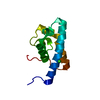
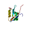
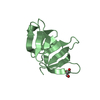
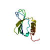
| Title | Crystal structure of the HRDC Domain of Human Werner Syndrome Protein, WRN | ||||||
 Components Components | Werner syndrome ATP-dependent helicase | ||||||
 Keywords Keywords | HYDROLASE / HRDC domain | ||||||
| Function / homology |  Function and homology information Function and homology information3'-flap-structured DNA binding / positive regulation of strand invasion / positive regulation of hydrolase activity / forked DNA-dependent helicase activity / telomeric G-quadruplex DNA binding / 8-hydroxy-2'-deoxyguanosine DNA binding / telomeric D-loop binding / regulation of growth rate / DNA geometric change / telomere maintenance via semi-conservative replication ...3'-flap-structured DNA binding / positive regulation of strand invasion / positive regulation of hydrolase activity / forked DNA-dependent helicase activity / telomeric G-quadruplex DNA binding / 8-hydroxy-2'-deoxyguanosine DNA binding / telomeric D-loop binding / regulation of growth rate / DNA geometric change / telomere maintenance via semi-conservative replication / Y-form DNA binding / telomeric D-loop disassembly / four-way junction helicase activity / t-circle formation / G-quadruplex DNA binding / bubble DNA binding / MutLalpha complex binding / Impaired BRCA2 binding to PALB2 / protein localization to nucleolus / Processive synthesis on the C-strand of the telomere / response to UV-C / Removal of the Flap Intermediate from the C-strand / exonuclease activity / HDR through Single Strand Annealing (SSA) / DNA metabolic process / Homologous DNA Pairing and Strand Exchange / Defective homologous recombination repair (HRR) due to BRCA1 loss of function / Defective HDR through Homologous Recombination Repair (HRR) due to PALB2 loss of BRCA1 binding function / Defective HDR through Homologous Recombination Repair (HRR) due to PALB2 loss of BRCA2/RAD51/RAD51C binding function / Resolution of D-loop Structures through Synthesis-Dependent Strand Annealing (SDSA) / DNA synthesis involved in DNA repair / Resolution of D-loop Structures through Holliday Junction Intermediates / 3'-5' DNA helicase activity / Impaired BRCA2 binding to RAD51 / DNA 3'-5' helicase / replication fork processing / replicative senescence / Presynaptic phase of homologous DNA pairing and strand exchange / mismatch repair / SUMOylation of DNA damage response and repair proteins / four-way junction DNA binding / 3'-5' exonuclease activity / telomere maintenance / cellular response to starvation / replication fork / DNA helicase activity / determination of adult lifespan / cellular response to gamma radiation / base-excision repair / G2/M DNA damage checkpoint / double-strand break repair via homologous recombination / HDR through Homologous Recombination (HRR) / cellular senescence / manganese ion binding / double-strand break repair / chromosome / Processing of DNA double-strand break ends / response to oxidative stress / Regulation of TP53 Activity through Phosphorylation / Hydrolases; Acting on ester bonds / DNA replication / chromosome, telomeric region / nuclear speck / DNA damage response / centrosome / protein-containing complex binding / nucleolus / magnesium ion binding / protein homodimerization activity / ATP hydrolysis activity / DNA binding / nucleoplasm / ATP binding / nucleus / cytoplasm Similarity search - Function | ||||||
| Biological species |  Homo sapiens (human) Homo sapiens (human) | ||||||
| Method |  X-RAY DIFFRACTION / SYNCHROTRON / MOLECULAR REPLACEMENT / Resolution: 2.3 Å X-RAY DIFFRACTION / SYNCHROTRON / MOLECULAR REPLACEMENT / Resolution: 2.3 Å | ||||||
 Authors Authors | Kitano, K. / Yoshihara, N. / Hakoshima, T. | ||||||
 Citation Citation | Journal: J.Biol.Chem. / Year: 2007 Title: Crystal structure of the HRDC domain of human Werner syndrome protein, WRN Authors: Kitano, K. / Yoshihara, N. / Hakoshima, T. | ||||||
| History |
|
- Structure visualization
| Structure viewer | Molecule: MolmilJmol/JSmol |
|---|
- Downloads & links
-Download
| PDBx/mmCIF format | 2e1e.cif.gz | 29.9 KB | Display | PDBx/mmCIF format |
|---|---|---|---|---|
| PDB format | pdb2e1e.ent.gz | 19.5 KB | Display | PDB format |
| PDBx/mmJSON format | 2e1e.json.gz | Tree view | PDBx/mmJSON format | |
| Others |  Other downloads Other downloads |
-Validation report
| Arichive directory | https://data.pdbj.org/pub/pdb/validation_reports/e1/2e1eftp://data.pdbj.org/pub/pdb/validation_reports/e1/2e1e | HTTPS FTP |
|---|
-Related structure data
| Related structure data |  2e1fC  1d8bS  1wudS C: citing same article ( S: Starting model for refinement |
|---|---|
| Similar structure data |


-Links
 PDBj
PDBj


- Assembly
| Deposited unit | 
| ||||||||
|---|---|---|---|---|---|---|---|---|---|
| 1 |
| ||||||||
| 2 | 
| ||||||||
| Unit cell |
| ||||||||
| Components on special symmetry positions |
|
-Components
| #1: Protein | Mass: 11399.173 Da / Num. of mol.: 1 / Fragment: HRDC domain Source method: isolated from a genetically manipulated source Source: (gene. exp.) Homo sapiens (human) / Plasmid: pGEX-5X-1 / Production host:  References: UniProt: Q14191, Hydrolases; Acting on acid anhydrides; In phosphorus-containing anhydrides |
|---|---|
| #2: Chemical | ChemComp-CL /   Mass: 35.453 Da / Num. of mol.: 1 / Source method: obtained synthetically / Formula: Cl Mass: 35.453 Da / Num. of mol.: 1 / Source method: obtained synthetically / Formula: Cl |
| #3: Water | ChemComp-HOH /  Mass: 18.015 Da / Num. of mol.: 29 / Source method: isolated from a natural source / Formula: H2O Mass: 18.015 Da / Num. of mol.: 29 / Source method: isolated from a natural source / Formula: H2O |
-Experimental details
-Experiment
| Experiment | Method: X-RAY DIFFRACTION / Number of used crystals: 1 |
|---|
- Sample preparation
| Crystal | Density Matthews: 4.32 Å3/Da / Density % sol: 71.52 % |
|---|---|
| Crystal grow | Temperature: 293 K / Method: vapor diffusion / pH: 9.5 / Details: pH 9.5, VAPOR DIFFUSION, temperature 293K |
-Data collection
| Diffraction | Mean temperature: 90 K |
|---|---|
| Diffraction source | Source: SYNCHROTRON / Site: SPring-8  / Beamline: BL38B1 / Wavelength: 1 Å / Beamline: BL38B1 / Wavelength: 1 Å |
| Detector | Type: RIGAKU / Detector: IMAGE PLATE / Date: Jul 1, 2006 |
| Radiation | Protocol: SINGLE WAVELENGTH / Monochromatic (M) / Laue (L): M / Scattering type: x-ray |
| Radiation wavelength | Wavelength: 1 Å / Relative weight: 1 |
| Reflection | Resolution: 2.3→20 Å / Num. obs: 9021 / % possible obs: 96.8 % |
| Reflection shell | Resolution: 2.3→2.38 Å / % possible all: 77.6 |
- Processing
| Software |
| ||||||||||||||||||||
|---|---|---|---|---|---|---|---|---|---|---|---|---|---|---|---|---|---|---|---|---|---|
| Refinement | Method to determine structure: MOLECULAR REPLACEMENT Starting model: 1D8B and 1WUD Resolution: 2.3→20 Å / Details: the structure was refined also with REFMAC 5.
| ||||||||||||||||||||
| Refinement step | Cycle: LAST / Resolution: 2.3→20 Å
|
