 Movie
Movie Controller


+ Open data

- Basic information
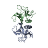
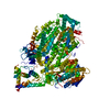
| Entry | Database: PDB / ID: 2dyn | ||||||
|---|---|---|---|---|---|---|---|
| Title | DYNAMIN (PLECKSTRIN HOMOLOGY DOMAIN) (DYNPH) | ||||||
 Components Components | DYNAMIN | ||||||
 Keywords Keywords | SIGNAL TRANSDUCTION / MOTOR PROTEIN / PHOSPHOLIPID BINDING / PROTEIN BINDING | ||||||
| Function / homology |  Function and homology information Function and homology informationclathrin coat assembly involved in endocytosis / vesicle scission / synaptic vesicle budding from presynaptic endocytic zone membrane / dynamin GTPase / chromaffin granule / regulation of vesicle size / Retrograde neurotrophin signalling / Toll Like Receptor 4 (TLR4) Cascade / Formation of annular gap junctions / endosome organization ...clathrin coat assembly involved in endocytosis / vesicle scission / synaptic vesicle budding from presynaptic endocytic zone membrane / dynamin GTPase / chromaffin granule / regulation of vesicle size / Retrograde neurotrophin signalling / Toll Like Receptor 4 (TLR4) Cascade / Formation of annular gap junctions / endosome organization / Gap junction degradation / Recycling pathway of L1 / phosphatidylinositol-3,4,5-trisphosphate binding / EPH-ephrin mediated repulsion of cells / endocytic vesicle / phosphatidylinositol-4,5-bisphosphate binding / clathrin-coated pit / MHC class II antigen presentation / receptor-mediated endocytosis / protein homooligomerization / endocytosis / GDP binding / Clathrin-mediated endocytosis / presynapse / microtubule binding / protein homotetramerization / microtubule / GTPase activity / synapse / protein kinase binding / GTP binding / protein homodimerization activity / RNA binding / extracellular exosome / identical protein binding / plasma membrane / cytoplasm Similarity search - Function | ||||||
| Biological species |  Homo sapiens (human) Homo sapiens (human) | ||||||
| Method |  X-RAY DIFFRACTION / SYNCHROTRON / MIR WITH ANOMALOUS SCATTERING / Resolution: 2.3 Å X-RAY DIFFRACTION / SYNCHROTRON / MIR WITH ANOMALOUS SCATTERING / Resolution: 2.3 Å | ||||||
 Authors Authors | Timm, D.E. | ||||||
 Citation Citation | Journal: Nat.Struct.Biol. / Year: 1994 Title: Crystal structure of the pleckstrin homology domain from dynamin. Authors: Timm, D. / Salim, K. / Gout, I. / Guruprasad, L. / Waterfield, M. / Blundell, T. | ||||||
| History |
|
- Structure visualization
| Structure viewer | Molecule: MolmilJmol/JSmol |
|---|
- Downloads & links
-Download
| PDBx/mmCIF format | 2dyn.cif.gz | 58.1 KB | Display | PDBx/mmCIF format |
|---|---|---|---|---|
| PDB format | pdb2dyn.ent.gz | 43.1 KB | Display | PDB format |
| PDBx/mmJSON format | 2dyn.json.gz | Tree view | PDBx/mmJSON format | |
| Others |  Other downloads Other downloads |
-Validation report
| Arichive directory | https://data.pdbj.org/pub/pdb/validation_reports/dy/2dynftp://data.pdbj.org/pub/pdb/validation_reports/dy/2dyn | HTTPS FTP |
|---|
-Related structure data
| Similar structure data |
|---|


-Links
 PDBj
PDBj


- Assembly
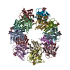
| Deposited unit | 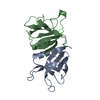
| ||||||||
|---|---|---|---|---|---|---|---|---|---|
| 1 | x 8
| ||||||||
| Unit cell |
| ||||||||
| Noncrystallographic symmetry (NCS) | NCS oper: (Code: given Matrix: (-0.847437, -0.116183, -0.518027), Vector: |
-Components
| #1: Protein | Mass: 14430.390 Da / Num. of mol.: 2 / Fragment: PLECKSTRIN HOMOLOGY (PH) DOMAIN Source method: isolated from a genetically manipulated source Source: (gene. exp.) Homo sapiens (human)Description: EXPRESSED AS GST-FUSION PROTEIN, CLEAVED WITH THROMBIN Plasmid: PGEX-2T / Production host:  #2: Water | ChemComp-HOH / |  Mass: 18.015 Da / Num. of mol.: 67 / Source method: isolated from a natural source / Formula: H2O Mass: 18.015 Da / Num. of mol.: 67 / Source method: isolated from a natural source / Formula: H2O |
|---|
-Experimental details
-Experiment
| Experiment | Method: X-RAY DIFFRACTION / Number of used crystals: 1 |
|---|
- Sample preparation
| Crystal | Density Matthews: 2.21 Å3/Da / Density % sol: 44.3 % | |||||||||||||||
|---|---|---|---|---|---|---|---|---|---|---|---|---|---|---|---|---|
| Crystal grow | pH: 6.1 Details: PROTEIN (10-20 MG/ML) WAS CRYSTALLIZED FROM 10-30% PEG 8000, 0.2 M AMMONIUM SULFATE, PH 6.1 BY THE HANGING DROP VAPOR DIFFUSION METHOD | |||||||||||||||
| Crystal grow | *PLUS Method: vapor diffusion, hanging drop | |||||||||||||||
| Components of the solutions | *PLUS
|
-Data collection
| Diffraction | Mean temperature: 298 K |
|---|---|
| Diffraction source | Source: SYNCHROTRON / Site: SRS  / Beamline: PX9.6 / Wavelength: 0.87 / Beamline: PX9.6 / Wavelength: 0.87 |
| Detector | Type: MARRESEARCH / Detector: IMAGE PLATE / Date: Jul 1, 1995 / Details: SYNCHROTRON |
| Radiation | Monochromator: SYNCHROTRON / Monochromatic (M) / Laue (L): M / Scattering type: x-ray |
| Radiation wavelength | Wavelength: 0.87 Å / Relative weight: 1 |
| Reflection | Resolution: 2.3→24.31 Å / Num. obs: 11759 / % possible obs: 99.5 % / Observed criterion σ(I): 2 / Redundancy: 6.6 % / Rmerge(I) obs: 0.058 / Rsym value: 0.058 / Net I/σ(I): 6.5 |
| Reflection shell | Resolution: 2.3→2.42 Å / Redundancy: 6.3 % / Rmerge(I) obs: 0.144 / Mean I/σ(I) obs: 5.1 / Rsym value: 0.144 / % possible all: 99.3 |
- Processing
| Software |
| ||||||||||||||||||||||||||||||||||||||||||||||||||||||||||||
|---|---|---|---|---|---|---|---|---|---|---|---|---|---|---|---|---|---|---|---|---|---|---|---|---|---|---|---|---|---|---|---|---|---|---|---|---|---|---|---|---|---|---|---|---|---|---|---|---|---|---|---|---|---|---|---|---|---|---|---|---|---|
| Refinement | Method to determine structure: MIR WITH ANOMALOUS SCATTERING Resolution: 2.3→24.31 Å / Data cutoff high absF: 100000 / Data cutoff low absF: 0.1 / Cross valid method: POSTERIORI / σ(F): 2 Details: A BULK SOLVENT MASK WAS APPLIED IN XPLOR 3.1. RESIDUES WITH OCCUPANCY OF 0.00 ARE INCLUDED IN THE MODEL, BUT ARE NOT DEFINED BY THE ELECTRON DENSITY.
| ||||||||||||||||||||||||||||||||||||||||||||||||||||||||||||
| Displacement parameters | Biso mean: 38.5 Å2 | ||||||||||||||||||||||||||||||||||||||||||||||||||||||||||||
| Refinement step | Cycle: LAST / Resolution: 2.3→24.31 Å
| ||||||||||||||||||||||||||||||||||||||||||||||||||||||||||||
| Refine LS restraints |
| ||||||||||||||||||||||||||||||||||||||||||||||||||||||||||||
| LS refinement shell | Resolution: 2.3→2.4 Å / Total num. of bins used: 8
| ||||||||||||||||||||||||||||||||||||||||||||||||||||||||||||
| Xplor file |
| ||||||||||||||||||||||||||||||||||||||||||||||||||||||||||||
| Software | *PLUS Name: X-PLOR / Version: 3.1 / Classification: refinement | ||||||||||||||||||||||||||||||||||||||||||||||||||||||||||||
| Refinement | *PLUS Rfactor Rfree: 0.2966 | ||||||||||||||||||||||||||||||||||||||||||||||||||||||||||||
| Solvent computation | *PLUS | ||||||||||||||||||||||||||||||||||||||||||||||||||||||||||||
| Displacement parameters | *PLUS | ||||||||||||||||||||||||||||||||||||||||||||||||||||||||||||
| Refine LS restraints | *PLUS
|
