 Movie
Movie Controller

[English] 日本語
 Yorodumi
- PDB-2du7: Crystal structure of Methanococcus jannacshii O-phosphoseryl-tRNA... -
+ Open data

- Basic information
| Entry | Database: PDB / ID: 2du7 | ||||||
|---|---|---|---|---|---|---|---|
| Title | Crystal structure of Methanococcus jannacshii O-phosphoseryl-tRNA synthetase | ||||||
 Components Components | O-phosphoseryl-tRNA synthetase | ||||||
 Keywords Keywords | LIGASE / alpha4 tetramer / Structural Genomics / NPPSFA / National Project on Protein Structural and Functional Analyses / RIKEN Structural Genomics/Proteomics Initiative / RSGI | ||||||
| Function / homology |  Function and homology information Function and homology informationO-phospho-L-serine-tRNA ligase / phosphoserine-tRNA(Cys) ligase activity / phenylalanyl-tRNA aminoacylation / phenylalanine-tRNA ligase activity / tRNA binding / ATP binding / cytoplasm Similarity search - Function | ||||||
| Biological species |   Methanocaldococcus jannaschii (archaea) Methanocaldococcus jannaschii (archaea) | ||||||
| Method |  X-RAY DIFFRACTION / SYNCHROTRON / MOLECULAR REPLACEMENT / Resolution: 3.6 Å X-RAY DIFFRACTION / SYNCHROTRON / MOLECULAR REPLACEMENT / Resolution: 3.6 Å | ||||||
 Authors Authors | Fukunaga, R. / RIKEN Structural Genomics/Proteomics Initiative (RSGI) | ||||||
 Citation Citation | Journal: Nat.Struct.Mol.Biol. / Year: 2007 Title: Structural insights into the first step of RNA-dependent cysteine biosynthesis in archaea. Authors: Fukunaga, R. / Yokoyama, S. | ||||||
| History |
|
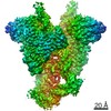
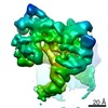
- Structure visualization
| Structure viewer | Molecule: MolmilJmol/JSmol |
|---|
- Downloads & links
-Download
| PDBx/mmCIF format | 2du7.cif.gz | 388.5 KB | Display | PDBx/mmCIF format |
|---|---|---|---|---|
| PDB format | pdb2du7.ent.gz | 322.7 KB | Display | PDB format |
| PDBx/mmJSON format | 2du7.json.gz | Tree view | PDBx/mmJSON format | |
| Others |  Other downloads Other downloads |
-Validation report
| Arichive directory | https://data.pdbj.org/pub/pdb/validation_reports/du/2du7ftp://data.pdbj.org/pub/pdb/validation_reports/du/2du7 | HTTPS FTP |
|---|
-Related structure data
| Related structure data |  2du3SC 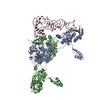 2du4C 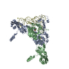 2du5C  2du6C S: Starting model for refinement C: citing same article ( |
|---|---|
| Similar structure data | |
| Other databases |
-Links
 PDBj
PDBj
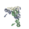
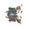
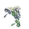
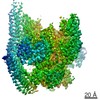
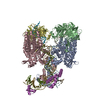
- Assembly
| Deposited unit | 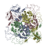
| ||||||||
|---|---|---|---|---|---|---|---|---|---|
| 1 |
| ||||||||
| 2 | 
| ||||||||
| Unit cell |
|
-Components
| #1: Protein | Mass: 63527.578 Da / Num. of mol.: 4 Source method: isolated from a genetically manipulated source Source: (gene. exp.) Methanocaldococcus jannaschii (archaea)Plasmid: pET26b / Production host:  References: UniProt: Q59054, Ligases; Forming carbon-oxygen bonds; Ligases forming aminoacyl-tRNA and related compounds |
|---|
-Experimental details
-Experiment
| Experiment | Method: X-RAY DIFFRACTION / Number of used crystals: 1 |
|---|
- Sample preparation
| Crystal | Density Matthews: 3.64 Å3/Da / Density % sol: 66.2 % |
|---|---|
| Crystal grow | Temperature: 293 K / Method: vapor diffusion, hanging drop / pH: 6.7 Details: 11.25% PEG 4000, 75mM Na-citrate, 75mM ADA-NaOH buffer (pH 6.7), VAPOR DIFFUSION, HANGING DROP, temperature 293K |
-Data collection
| Diffraction | Mean temperature: 173 K |
|---|---|
| Diffraction source | Source: SYNCHROTRON / Site: SPring-8  / Beamline: BL41XU / Wavelength: 1 Å / Beamline: BL41XU / Wavelength: 1 Å |
| Detector | Type: ADSC QUANTUM 315 / Detector: CCD / Date: Jun 30, 2005 |
| Radiation | Protocol: SINGLE WAVELENGTH / Monochromatic (M) / Laue (L): M / Scattering type: x-ray |
| Radiation wavelength | Wavelength: 1 Å / Relative weight: 1 |
| Reflection | Resolution: 3.6→20 Å / Num. all: 43084 / Num. obs: 42030 / % possible obs: 97.6 % / Observed criterion σ(I): 0 |
| Reflection shell | Resolution: 3.6→3.73 Å / % possible all: 94.1 |
- Processing
| Software |
| ||||||||||||||||||||||||||||||||||||
|---|---|---|---|---|---|---|---|---|---|---|---|---|---|---|---|---|---|---|---|---|---|---|---|---|---|---|---|---|---|---|---|---|---|---|---|---|---|
| Refinement | Method to determine structure: MOLECULAR REPLACEMENT Starting model: 2DU3 Resolution: 3.6→19.79 Å / Rfactor Rfree error: 0.006 / Data cutoff high absF: 7089748.71 / Data cutoff low absF: 0 / Isotropic thermal model: RESTRAINED / Cross valid method: THROUGHOUT / σ(F): 0
| ||||||||||||||||||||||||||||||||||||
| Solvent computation | Solvent model: FLAT MODEL / Bsol: 32.9985 Å2 / ksol: 0.194656 e/Å3 | ||||||||||||||||||||||||||||||||||||
| Displacement parameters | Biso mean: 129.2 Å2
| ||||||||||||||||||||||||||||||||||||
| Refine analyze |
| ||||||||||||||||||||||||||||||||||||
| Refinement step | Cycle: LAST / Resolution: 3.6→19.79 Å
| ||||||||||||||||||||||||||||||||||||
| Refine LS restraints |
| ||||||||||||||||||||||||||||||||||||
| LS refinement shell | Resolution: 3.6→3.82 Å / Rfactor Rfree error: 0.017 / Total num. of bins used: 6
| ||||||||||||||||||||||||||||||||||||
| Xplor file |
|
