 Movie
Movie Controller

[English] 日本語
 Yorodumi
- PDB-2drs: Crystal structure of reducing-end-xylose releasing exo-oligoxylan... -
+ Open data

- Basic information
| Entry | Database: PDB / ID: 2drs | ||||||
|---|---|---|---|---|---|---|---|
| Title | Crystal structure of reducing-end-xylose releasing exo-oligoxylanase D263S mutant | ||||||
 Components Components | Xylanase Y | ||||||
 Keywords Keywords | HYDROLASE / (ALPLA/ALPHA)6 BARREL / GLYCOSIDE HYDROLASE FAMILY 8 / Structural Genomics / NPPSFA / National Project on Protein Structural and Functional Analyses | ||||||
| Function / homology |  Function and homology information Function and homology informationoligosaccharide reducing-end xylanase / oligosaccharide reducing-end xylanase activity / xylan catabolic process Similarity search - Function | ||||||
| Biological species |  Bacillus halodurans (bacteria) Bacillus halodurans (bacteria) | ||||||
| Method |  X-RAY DIFFRACTION / SYNCHROTRON / FOURIER SYNTHESIS / Resolution: 2.1 Å X-RAY DIFFRACTION / SYNCHROTRON / FOURIER SYNTHESIS / Resolution: 2.1 Å | ||||||
 Authors Authors | Fushinobu, S. / Hidaka, M. / Honda, Y. / Wakagi, T. / Shoun, H. / Kitaoka, M. | ||||||
 Citation Citation | Journal: J.Biochem. / Year: 2009 Title: Structural explanation for the acquisition of glycosynthase activity Authors: Hidaka, M. / Fushinobu, S. / Honda, Y. / Wakagi, T. / Shoun, H. / Kitaoka, M. #1: Journal: J.Biol.Chem. / Year: 2006 Title: The first glycosynthase derived from an inverting glycoside hydrolase Authors: Honda, Y. / Kitaoka, M. #2: Journal: J.Biol.Chem. / Year: 2005Title: Structural basis for the specificity of the reducing end xylose-releasing exo-oligoxylanase from Bacillus halodurans C-125 Authors: Fushinobu, S. / Hidaka, M. / Honda, Y. / Wakagi, T. / Shoun, H. / Kitaoka, M. #3: Journal: Acta Crystallogr.,Sect.F / Year: 2005 Title: Crystallization and preliminary X-ray analysis of reducing-end xylose-releasing exo-oligoxylanase from Bacillus halodurans C-125 Authors: Honda, Y. / Fushinobu, S. / Hidaka, M. / Wakagi, T. / Shoun, H. / Kitaoka, M. #4: Journal: J.Biol.Chem. / Year: 2004 Title: A family 8 glycoside hydrolase from Bacillus halodurans C-125 (BH2105) is a reducing end xylose-releasing exo-oligoxylanase Authors: Honda, Y. / Kitaoka, M. | ||||||
| History |
|
- Structure visualization
| Structure viewer | Molecule: MolmilJmol/JSmol |
|---|
- Downloads & links
-Download
| PDBx/mmCIF format | 2drs.cif.gz | 100.6 KB | Display | PDBx/mmCIF format |
|---|---|---|---|---|
| PDB format | pdb2drs.ent.gz | 75.6 KB | Display | PDB format |
| PDBx/mmJSON format | 2drs.json.gz | Tree view | PDBx/mmJSON format | |
| Others |  Other downloads Other downloads |
-Validation report
| Arichive directory | https://data.pdbj.org/pub/pdb/validation_reports/dr/2drsftp://data.pdbj.org/pub/pdb/validation_reports/dr/2drs | HTTPS FTP |
|---|
-Related structure data
| Related structure data | 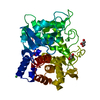 2droC 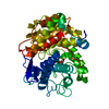 2drqC 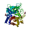 2drrC 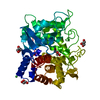 3a3vC  1wu4S S: Starting model for refinement C: citing same article ( |
|---|---|
| Similar structure data |


-Links
 PDBj
PDBj
- Assembly
| Deposited unit | 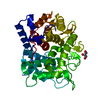
| ||||||||
|---|---|---|---|---|---|---|---|---|---|
| 1 |
| ||||||||
| Unit cell |
|
-Components
| #1: Protein | Mass: 46100.793 Da / Num. of mol.: 1 / Mutation: K2E/D263S Source method: isolated from a genetically manipulated source Source: (gene. exp.) Bacillus halodurans (bacteria) / Strain: C-125 / Gene: BH2105 / Plasmid: PET28B-BH2105 / Production host: References: UniProt: Q9KB30, oligosaccharide reducing-end xylanase |
|---|---|
| #2: Chemical | ChemComp-NI /   Mass: 58.693 Da / Num. of mol.: 1 / Source method: obtained synthetically / Formula: Ni Mass: 58.693 Da / Num. of mol.: 1 / Source method: obtained synthetically / Formula: Ni |
| #3: Chemical | ChemComp-GOL /   Mass: 92.094 Da / Num. of mol.: 1 / Source method: obtained synthetically / Formula: C3H8O3 Mass: 92.094 Da / Num. of mol.: 1 / Source method: obtained synthetically / Formula: C3H8O3 |
| #4: Water | ChemComp-HOH /  Mass: 18.015 Da / Num. of mol.: 431 / Source method: isolated from a natural source / Formula: H2O Mass: 18.015 Da / Num. of mol.: 431 / Source method: isolated from a natural source / Formula: H2O |
-Experimental details
-Experiment
| Experiment | Method: X-RAY DIFFRACTION / Number of used crystals: 1 |
|---|
- Sample preparation
| Crystal | Density Matthews: 2.09 Å3/Da / Density % sol: 41.17 % |
|---|---|
| Crystal grow | Temperature: 293 K / Method: vapor diffusion, hanging drop / pH: 4.6 Details: PEG4000, SODIUM ACETATE, GLYCEROL, pH 4.6, VAPOR DIFFUSION, HANGING DROP, temperature 293K |
-Data collection
| Diffraction | Mean temperature: 100 K |
|---|---|
| Diffraction source | Source: SYNCHROTRON / Site: Photon Factory  / Beamline: BL-6A / Wavelength: 1 Å / Beamline: BL-6A / Wavelength: 1 Å |
| Detector | Type: ADSC QUAMTUM 4r / Detector: CCD / Date: Oct 22, 2004 |
| Radiation | Monochromator: Triangular Si(111) with an asymmetric angle of 7.8degree Protocol: SINGLE WAVELENGTH / Monochromatic (M) / Laue (L): M / Scattering type: x-ray |
| Radiation wavelength | Wavelength: 1 Å / Relative weight: 1 |
| Reflection | Resolution: 2.1→60.746 Å / Num. all: 23314 / Num. obs: 22673 / % possible obs: 97.3 % / Observed criterion σ(F): 0 / Observed criterion σ(I): 0 / Redundancy: 4.5 % / Biso Wilson estimate: 12.8 Å2 / Rsym value: 0.099 / Net I/σ(I): 15.2 |
| Reflection shell | Resolution: 2.1→2.18 Å / Redundancy: 4.5 % / Mean I/σ(I) obs: 5.9 / Num. unique all: 2294 / Rsym value: 0.243 / % possible all: 99.2 |
- Processing
| Software |
| ||||||||||||||||||||||||||||||||||||||||||||||||||||||||||||||||||||||||||||||||||||||||||||||||||||||||||||||||||||||||||||||||||||||||||||||||||||||||||||||||||||||||||
|---|---|---|---|---|---|---|---|---|---|---|---|---|---|---|---|---|---|---|---|---|---|---|---|---|---|---|---|---|---|---|---|---|---|---|---|---|---|---|---|---|---|---|---|---|---|---|---|---|---|---|---|---|---|---|---|---|---|---|---|---|---|---|---|---|---|---|---|---|---|---|---|---|---|---|---|---|---|---|---|---|---|---|---|---|---|---|---|---|---|---|---|---|---|---|---|---|---|---|---|---|---|---|---|---|---|---|---|---|---|---|---|---|---|---|---|---|---|---|---|---|---|---|---|---|---|---|---|---|---|---|---|---|---|---|---|---|---|---|---|---|---|---|---|---|---|---|---|---|---|---|---|---|---|---|---|---|---|---|---|---|---|---|---|---|---|---|---|---|---|---|---|
| Refinement | Method to determine structure: FOURIER SYNTHESIS Starting model: PDB ENTRY 1WU4 Resolution: 2.1→43.4 Å / Cor.coef. Fo:Fc: 0.956 / Cor.coef. Fo:Fc free: 0.91 / SU B: 3.956 / SU ML: 0.109 / Isotropic thermal model: Isotropic / Cross valid method: THROUGHOUT / σ(F): 0 / ESU R: 0.21 / ESU R Free: 0.184 / Stereochemistry target values: MAXIMUM LIKELIHOOD / Details: HYDROGENS HAVE BEEN ADDED IN THE RIDING POSITIONS
| ||||||||||||||||||||||||||||||||||||||||||||||||||||||||||||||||||||||||||||||||||||||||||||||||||||||||||||||||||||||||||||||||||||||||||||||||||||||||||||||||||||||||||
| Solvent computation | Ion probe radii: 0.8 Å / Shrinkage radii: 0.8 Å / VDW probe radii: 1.4 Å / Solvent model: MASK | ||||||||||||||||||||||||||||||||||||||||||||||||||||||||||||||||||||||||||||||||||||||||||||||||||||||||||||||||||||||||||||||||||||||||||||||||||||||||||||||||||||||||||
| Displacement parameters | Biso mean: 10.255 Å2
| ||||||||||||||||||||||||||||||||||||||||||||||||||||||||||||||||||||||||||||||||||||||||||||||||||||||||||||||||||||||||||||||||||||||||||||||||||||||||||||||||||||||||||
| Refine analyze | Luzzati coordinate error obs: 0.1788 Å | ||||||||||||||||||||||||||||||||||||||||||||||||||||||||||||||||||||||||||||||||||||||||||||||||||||||||||||||||||||||||||||||||||||||||||||||||||||||||||||||||||||||||||
| Refinement step | Cycle: LAST / Resolution: 2.1→43.4 Å
| ||||||||||||||||||||||||||||||||||||||||||||||||||||||||||||||||||||||||||||||||||||||||||||||||||||||||||||||||||||||||||||||||||||||||||||||||||||||||||||||||||||||||||
| Refine LS restraints |
| ||||||||||||||||||||||||||||||||||||||||||||||||||||||||||||||||||||||||||||||||||||||||||||||||||||||||||||||||||||||||||||||||||||||||||||||||||||||||||||||||||||||||||
| LS refinement shell | Resolution: 2.098→2.153 Å / Total num. of bins used: 20
|
