 Movie
Movie Controller

[English] 日本語
 Yorodumi
- PDB-3a3v: Crystal structure of reducing-end-xylose releasing exo-oligoxylan... -
+ Open data

- Basic information
| Entry | Database: PDB / ID: 3a3v | ||||||
|---|---|---|---|---|---|---|---|
| Title | Crystal structure of reducing-end-xylose releasing exo-oligoxylanase Y198F mutant | ||||||
 Components Components | Xylanase Y | ||||||
 Keywords Keywords | HYDROLASE / Xylan degradation | ||||||
| Function / homology |  Function and homology information Function and homology informationoligosaccharide reducing-end xylanase / oligosaccharide reducing-end xylanase activity / xylan catabolic process Similarity search - Function | ||||||
| Biological species |  Bacillus halodurans (bacteria) Bacillus halodurans (bacteria) | ||||||
| Method |  X-RAY DIFFRACTION / SYNCHROTRON / MOLECULAR REPLACEMENT / molecular replacement / Resolution: 1.39 Å X-RAY DIFFRACTION / SYNCHROTRON / MOLECULAR REPLACEMENT / molecular replacement / Resolution: 1.39 Å | ||||||
 Authors Authors | Hidaka, M. / Fushinobu, S. / Honda, Y. / Kitaoka, M. | ||||||
 Citation Citation | Journal: J.Biochem. / Year: 2010 Title: Structural explanation for the acquisition of glycosynthase activity Authors: Hidaka, M. / Fushinobu, S. / Honda, Y. / Wakagi, T. / Shoun, H. / Kitaoka, M. #1: Journal: Glycobiology / Year: 2008 Title: Alternative strategy for converting an inverting glycoside hydrolase into a glycosynthase Authors: Honda, Y. / Fushinobu, S. / Hidaka, M. / Wakagi, T. / Shoun, H. / Taniguchi, H. / Kitaoka, M. #2: Journal: J.Biol.Chem. / Year: 2006 Title: The first glycosynthase derived from an inverting glycoside hydrolase Authors: Honda, Y. / Kitaoka, M. #3: Journal: J.Biol.Chem. / Year: 2005Title: Structural basis for the specificity of the reducing end xylose-releasing exo-oligoxylanase from Bacillus halodurans C-125 Authors: Fushinobu, S. / Hidaka, M. / Honda, Y. / Wakagi, T. / Shoun, H. / Kitaoka, M. #4: Journal: Acta Crystallogr.,Sect.F / Year: 2005 Title: Crystallization and preliminary X-ray analysis of reducing-end xylose-releasing exo-oligoxylanase from Bacillus halodurans C-125 Authors: Honda, Y. / Fushinobu, S. / Hidaka, M. / Wakagi, T. / Shoun, H. / Kitaoka, M. #5: Journal: J.Biol.Chem. / Year: 2004 Title: A family 8 glycoside hydrolase from Bacillus halodurans C-125 (BH2105) is a reducing end xylose-releasing exo-oligoxylanase Authors: Honda, Y. / Kitaoka, M. | ||||||
| History |
|
- Structure visualization
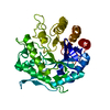
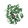
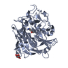
| Structure viewer | Molecule: MolmilJmol/JSmol |
|---|
- Downloads & links
-Download
| PDBx/mmCIF format | 3a3v.cif.gz | 103.2 KB | Display | PDBx/mmCIF format |
|---|---|---|---|---|
| PDB format | pdb3a3v.ent.gz | 77.3 KB | Display | PDB format |
| PDBx/mmJSON format | 3a3v.json.gz | Tree view | PDBx/mmJSON format | |
| Others |  Other downloads Other downloads |
-Validation report
| Arichive directory | https://data.pdbj.org/pub/pdb/validation_reports/a3/3a3vftp://data.pdbj.org/pub/pdb/validation_reports/a3/3a3v | HTTPS FTP |
|---|
-Related structure data
| Related structure data | 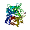 2droC 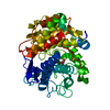 2drqC 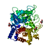 2drrC  2drsC 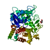 1wu4S S: Starting model for refinement C: citing same article ( |
|---|---|
| Similar structure data |


-Links
 PDBj
PDBj
- Assembly
| Deposited unit | 
| ||||||||
|---|---|---|---|---|---|---|---|---|---|
| 1 |
| ||||||||
| Unit cell |
|
-Components
| #1: Protein | Mass: 46112.801 Da / Num. of mol.: 1 / Mutation: K2E,Y198F Source method: isolated from a genetically manipulated source Source: (gene. exp.) Bacillus halodurans (bacteria) / Strain: C-125 / Gene: BH2105 / Plasmid: PET28B-BH2105 / Production host: References: UniProt: Q9KB30, oligosaccharide reducing-end xylanase | ||
|---|---|---|---|
| #2: Chemical | ChemComp-NI /   Mass: 58.693 Da / Num. of mol.: 1 / Source method: obtained synthetically / Formula: Ni Mass: 58.693 Da / Num. of mol.: 1 / Source method: obtained synthetically / Formula: Ni | ||
| #3: Chemical | ChemComp-GOL /   Mass: 92.094 Da / Num. of mol.: 4 / Source method: obtained synthetically / Formula: C3H8O3 Mass: 92.094 Da / Num. of mol.: 4 / Source method: obtained synthetically / Formula: C3H8O3#4: Water | ChemComp-HOH / |  Mass: 18.015 Da / Num. of mol.: 516 / Source method: isolated from a natural source / Formula: H2O Mass: 18.015 Da / Num. of mol.: 516 / Source method: isolated from a natural source / Formula: H2O |
-Experimental details
-Experiment
| Experiment | Method: X-RAY DIFFRACTION / Number of used crystals: 1 |
|---|
- Sample preparation
| Crystal | Density Matthews: 2.18 Å3/Da / Density % sol: 43.6 % |
|---|---|
| Crystal grow | Temperature: 293 K / Method: vapor diffusion, hanging drop / pH: 4.6 Details: PEG4000, SODIUM ACETATE, GLYCEROL, pH 4.6, VAPOR DIFFUSION, HANGING DROP, temperature 293K |
-Data collection
| Diffraction | Mean temperature: 95 K |
|---|---|
| Diffraction source | Source: SYNCHROTRON / Site: Photon Factory  / Beamline: BL-17A / Wavelength: 1 Å / Beamline: BL-17A / Wavelength: 1 Å |
| Detector | Type: ADSC QUANTUM 4 / Detector: CCD / Date: Mar 11, 2007 |
| Radiation | Monochromator: Numerical link type Si(111)double crystal monochromator Protocol: SINGLE WAVELENGTH / Monochromatic (M) / Laue (L): M / Scattering type: x-ray |
| Radiation wavelength | Wavelength: 1 Å / Relative weight: 1 |
| Reflection | Resolution: 1.39→50 Å / Num. all: 81959 / Num. obs: 81761 / % possible obs: 99.7 % / Redundancy: 3.7 % / Biso Wilson estimate: 19.7 Å2 / Rmerge(I) obs: 0.079 / Net I/σ(I): 32.1 |
| Reflection shell | Resolution: 1.39→1.44 Å / Redundancy: 3.4 % / Rmerge(I) obs: 0.384 / Mean I/σ(I) obs: 3.5 / % possible all: 97.6 |
-Phasing
| Phasing | Method: molecular replacement |
|---|
- Processing
| Software |
| |||||||||||||||||||||||||||||||||||||||||||||||||||||||||||||||||
|---|---|---|---|---|---|---|---|---|---|---|---|---|---|---|---|---|---|---|---|---|---|---|---|---|---|---|---|---|---|---|---|---|---|---|---|---|---|---|---|---|---|---|---|---|---|---|---|---|---|---|---|---|---|---|---|---|---|---|---|---|---|---|---|---|---|---|
| Refinement | Method to determine structure: MOLECULAR REPLACEMENT Starting model: 1WU4 Resolution: 1.39→45.5 Å / Cor.coef. Fo:Fc: 0.964 / Cor.coef. Fo:Fc free: 0.953 / WRfactor Rfree: 0.19 / WRfactor Rwork: 0.17 / Occupancy max: 1 / Occupancy min: 1 / FOM work R set: 0.888 / SU R Cruickshank DPI: 0.06 / SU Rfree: 0.061 / Cross valid method: THROUGHOUT / σ(F): 0 / ESU R: 0.06 / ESU R Free: 0.061 / Stereochemistry target values: MAXIMUM LIKELIHOOD Details: HYDROGENS HAVE BEEN ADDED IN THE RIDING POSITIONS U VALUES: REFINED INDIVIDUALLY
| |||||||||||||||||||||||||||||||||||||||||||||||||||||||||||||||||
| Solvent computation | Ion probe radii: 0.8 Å / Shrinkage radii: 0.8 Å / VDW probe radii: 1.4 Å / Solvent model: MASK | |||||||||||||||||||||||||||||||||||||||||||||||||||||||||||||||||
| Displacement parameters | Biso max: 50.25 Å2 / Biso mean: 14.73 Å2 / Biso min: 5.92 Å2
| |||||||||||||||||||||||||||||||||||||||||||||||||||||||||||||||||
| Refine analyze | Luzzati coordinate error obs: 0.1439 Å | |||||||||||||||||||||||||||||||||||||||||||||||||||||||||||||||||
| Refinement step | Cycle: LAST / Resolution: 1.39→45.5 Å
| |||||||||||||||||||||||||||||||||||||||||||||||||||||||||||||||||
| Refine LS restraints |
| |||||||||||||||||||||||||||||||||||||||||||||||||||||||||||||||||
| LS refinement shell | Resolution: 1.39→1.426 Å / Total num. of bins used: 20
|
