 Movie
Movie Controller

[English] 日本語
 Yorodumi
- PDB-1wu6: Crystal structure of reducing-end-xylose releasing exo-oligoxylan... -
+ Open data

- Basic information
| Entry | Database: PDB / ID: 1wu6 | |||||||||
|---|---|---|---|---|---|---|---|---|---|---|
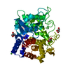
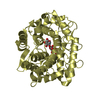
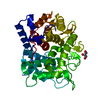
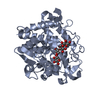
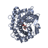
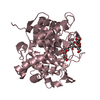
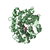
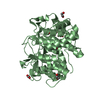
| Title | Crystal structure of reducing-end-xylose releasing exo-oligoxylanase E70A mutant complexed with xylobiose | |||||||||
 Components Components | xylanase Y | |||||||||
 Keywords Keywords | HYDROLASE / (alpla/alpha)6 barrel / glycoside hydrolase family 8 | |||||||||
| Function / homology |  Function and homology information Function and homology informationoligosaccharide reducing-end xylanase / oligosaccharide reducing-end xylanase activity / xylan catabolic process Similarity search - Function | |||||||||
| Biological species |  Bacillus halodurans (bacteria) Bacillus halodurans (bacteria) | |||||||||
| Method |  X-RAY DIFFRACTION / SYNCHROTRON / MOLECULAR REPLACEMENT / Resolution: 1.45 Å X-RAY DIFFRACTION / SYNCHROTRON / MOLECULAR REPLACEMENT / Resolution: 1.45 Å | |||||||||
 Authors Authors | Fushinobu, S. / Hidaka, M. / Honda, Y. / Wakagi, T. / Shoun, H. / Kitaoka, M. | |||||||||
 Citation Citation | Journal: J.Biol.Chem. / Year: 2005 Title: Structural Basis for the Specificity of the Reducing End Xylose-releasing Exo-oligoxylanase from Bacillus halodurans C-125 Authors: Fushinobu, S. / Hidaka, M. / Honda, Y. / Wakagi, T. / Shoun, H. / Kitaoka, M. #1: Journal: To be PublishedTitle: Crystallization and preliminary X-ray analysis of reducing-end-xylose releasing exo-oligoxylanase (Rex) form Bacillus halodurans C-125 Authors: Honda, Y. / Fushinobu, S. / Hidaka, M. / Wakagi, T. / Shoun, H. / Kitaoka, M. #2: Journal: J.Biol.Chem. / Year: 2004 Title: A family 8 glycoside hydrolase from Bacillus halodurans C-125 (BH2105) is a reducing end xylose-releasing exo-oligoxylanase Authors: Honda, Y. / Kitaoka, M. | |||||||||
| History |
|
- Structure visualization
| Structure viewer | Molecule: MolmilJmol/JSmol |
|---|
- Downloads & links
-Download
| PDBx/mmCIF format | 1wu6.cif.gz | 103.3 KB | Display | PDBx/mmCIF format |
|---|---|---|---|---|
| PDB format | pdb1wu6.ent.gz | 76.4 KB | Display | PDB format |
| PDBx/mmJSON format | 1wu6.json.gz | Tree view | PDBx/mmJSON format | |
| Others |  Other downloads Other downloads |
-Validation report
| Arichive directory | https://data.pdbj.org/pub/pdb/validation_reports/wu/1wu6ftp://data.pdbj.org/pub/pdb/validation_reports/wu/1wu6 | HTTPS FTP |
|---|
-Related structure data
| Related structure data | 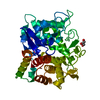 1wu4C 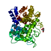 1wu5C 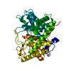 1h14S C: citing same article ( S: Starting model for refinement |
|---|---|
| Similar structure data |


-Links
 PDBj
PDBj
- Assembly
| Deposited unit | 
| ||||||||
|---|---|---|---|---|---|---|---|---|---|
| 1 |
| ||||||||
| Unit cell |
| ||||||||
| Details | The biological assembly is monomer in the asymmetric unit. |
-Components
| #1: Protein | Mass: 46070.766 Da / Num. of mol.: 1 / Mutation: E70A Source method: isolated from a genetically manipulated source Source: (gene. exp.) Bacillus halodurans (bacteria) / Strain: C-125 / Gene: BH2105 / Plasmid: pET28b-BH2105 / Production host: References: GenBank: 15614668, UniProt: Q9KB30*PLUS, oligosaccharide reducing-end xylanase | ||
|---|---|---|---|
| #2: Polysaccharide | beta-D-xylopyranose-(1-4)-beta-D-xylopyranose / 4beta-beta-xylobiose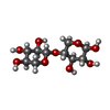  Source method: isolated from a genetically manipulated source Details: oligosaccharide / References: 4beta-beta-xylobiose | ||
| #3: Chemical | ChemComp-NI /   Mass: 58.693 Da / Num. of mol.: 1 / Source method: obtained synthetically / Formula: Ni Mass: 58.693 Da / Num. of mol.: 1 / Source method: obtained synthetically / Formula: Ni | ||
| #4: Chemical |   Mass: 92.094 Da / Num. of mol.: 2 / Source method: obtained synthetically / Formula: C3H8O3 Mass: 92.094 Da / Num. of mol.: 2 / Source method: obtained synthetically / Formula: C3H8O3#5: Water | ChemComp-HOH / |  Mass: 18.015 Da / Num. of mol.: 492 / Source method: isolated from a natural source / Formula: H2O Mass: 18.015 Da / Num. of mol.: 492 / Source method: isolated from a natural source / Formula: H2O |
-Experimental details
-Experiment
| Experiment | Method: X-RAY DIFFRACTION / Number of used crystals: 1 |
|---|
- Sample preparation
| Crystal | Density Matthews: 2.21 Å3/Da / Density % sol: 44.3 % |
|---|---|
| Crystal grow | Temperature: 293 K / Method: vapor diffusion, hanging drop / pH: 4.6 Details: PEG4000, sodium acetate, glycerol, pH 4.6, VAPOR DIFFUSION, HANGING DROP, temperature 293K |
-Data collection
| Diffraction | Mean temperature: 100 K |
|---|---|
| Diffraction source | Source: SYNCHROTRON / Site: Photon Factory  / Beamline: AR-NW12A / Wavelength: 1 Å / Beamline: AR-NW12A / Wavelength: 1 Å |
| Detector | Type: ADSC QUANTUM 210 / Detector: CCD / Date: Jun 30, 2004 |
| Radiation | Monochromator: Si(111) double crystal / Protocol: SINGLE WAVELENGTH / Monochromatic (M) / Laue (L): M / Scattering type: x-ray |
| Radiation wavelength | Wavelength: 1 Å / Relative weight: 1 |
| Reflection | Resolution: 1.45→62.02 Å / Num. all: 72773 / Num. obs: 72191 / % possible obs: 99.2 % / Observed criterion σ(F): 0 / Observed criterion σ(I): 0 / Biso Wilson estimate: 13.1 Å2 / Rsym value: 0.053 / Net I/σ(I): 26.5 |
| Reflection shell | Resolution: 1.45→1.5 Å / Mean I/σ(I) obs: 3.9 / Rsym value: 0.295 / % possible all: 97.9 |
- Processing
| Software |
| ||||||||||||||||||||||||||||||||||||||||||||||||||||||||||||||||||||||||||||||||
|---|---|---|---|---|---|---|---|---|---|---|---|---|---|---|---|---|---|---|---|---|---|---|---|---|---|---|---|---|---|---|---|---|---|---|---|---|---|---|---|---|---|---|---|---|---|---|---|---|---|---|---|---|---|---|---|---|---|---|---|---|---|---|---|---|---|---|---|---|---|---|---|---|---|---|---|---|---|---|---|---|---|
| Refinement | Method to determine structure: MOLECULAR REPLACEMENT Starting model: PDB ENTRY 1H14 Resolution: 1.45→31.39 Å / Rfactor Rfree error: 0.003 / Data cutoff high absF: 1578112.8 / Data cutoff low absF: 0 / Isotropic thermal model: RESTRAINED / Cross valid method: THROUGHOUT / σ(F): 0 / Stereochemistry target values: Engh & Huber
| ||||||||||||||||||||||||||||||||||||||||||||||||||||||||||||||||||||||||||||||||
| Solvent computation | Solvent model: FLAT MODEL / Bsol: 55.8997 Å2 / ksol: 0.413878 e/Å3 | ||||||||||||||||||||||||||||||||||||||||||||||||||||||||||||||||||||||||||||||||
| Displacement parameters | Biso mean: 14.9 Å2
| ||||||||||||||||||||||||||||||||||||||||||||||||||||||||||||||||||||||||||||||||
| Refine analyze |
| ||||||||||||||||||||||||||||||||||||||||||||||||||||||||||||||||||||||||||||||||
| Refinement step | Cycle: LAST / Resolution: 1.45→31.39 Å
| ||||||||||||||||||||||||||||||||||||||||||||||||||||||||||||||||||||||||||||||||
| Refine LS restraints |
| ||||||||||||||||||||||||||||||||||||||||||||||||||||||||||||||||||||||||||||||||
| LS refinement shell | Resolution: 1.45→1.54 Å / Rfactor Rfree error: 0.009 / Total num. of bins used: 6
| ||||||||||||||||||||||||||||||||||||||||||||||||||||||||||||||||||||||||||||||||
| Xplor file |
|
