 Movie
Movie Controller

[English] 日本語
 Yorodumi
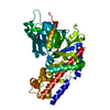
- PDB-2dg6: Crystal structure of the putative transcriptional regulator SCO55... -
+ Open data

- Basic information
| Entry | Database: PDB / ID: 2dg6 | ||||||
|---|---|---|---|---|---|---|---|
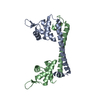
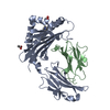
| Title | Crystal structure of the putative transcriptional regulator SCO5550 from Streptomyces coelicolor A3(2) | ||||||
 Components Components | putative transcriptional regulator | ||||||
 Keywords Keywords | GENE REGULATION / Winged-helix motif / MerR family | ||||||
| Function / homology |  Function and homology information Function and homology information | ||||||
| Biological species |  Streptomyces coelicolor (bacteria) Streptomyces coelicolor (bacteria) | ||||||
| Method |  X-RAY DIFFRACTION / SYNCHROTRON / MAD / Resolution: 2.2 Å X-RAY DIFFRACTION / SYNCHROTRON / MAD / Resolution: 2.2 Å | ||||||
 Authors Authors | Hayashi, T. / Tanaka, Y. / Sakai, N. / Yao, M. / Tamura, T. / Tanaka, I. | ||||||
 Citation Citation | Journal: Biochem. Biophys. Res. Commun. / Year: 2013 Title: Structural and genomic DNA analysis of a putative transcription factor SCO5550 from Streptomyces coelicolor A3(2): regulating the expression of gene sco5551 as a transcriptional activator with a novel dimer shape Authors: Hayashi, T. / Tanaka, Y. / Sakai, N. / Watanabe, N. / Tamura, T. / Tanaka, I. / Yao, M. | ||||||
| History |
|
- Structure visualization
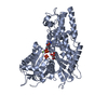
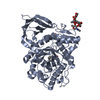
| Structure viewer | Molecule: MolmilJmol/JSmol |
|---|
- Downloads & links
-Download
| PDBx/mmCIF format | 2dg6.cif.gz | 50.8 KB | Display | PDBx/mmCIF format |
|---|---|---|---|---|
| PDB format | pdb2dg6.ent.gz | 39.7 KB | Display | PDB format |
| PDBx/mmJSON format | 2dg6.json.gz | Tree view | PDBx/mmJSON format | |
| Others |  Other downloads Other downloads |
-Validation report
| Summary document | 2dg6_validation.pdf.gz | 425.3 KB | Display | wwPDB validaton report |
|---|---|---|---|---|
| Full document | 2dg6_full_validation.pdf.gz | 428.9 KB | Display | |
| Data in XML | 2dg6_validation.xml.gz | 10.6 KB | Display | |
| Data in CIF | 2dg6_validation.cif.gz | 14.2 KB | Display | |
| Arichive directory | https://data.pdbj.org/pub/pdb/validation_reports/dg/2dg6ftp://data.pdbj.org/pub/pdb/validation_reports/dg/2dg6 | HTTPS FTP |
-Related structure data
| Related structure data | |
|---|---|
| Similar structure data |


-Links
 PDBj
PDBj

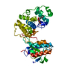
- Assembly
| Deposited unit | 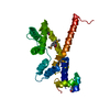
| ||||||||
|---|---|---|---|---|---|---|---|---|---|
| 1 | 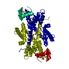
| ||||||||
| Unit cell |
| ||||||||
| Details | The biological assembly is a dimer generated from the monomer in the asymmetric unit by the operations: -x, -y+1, z. |
-Components
| #1: Protein | Mass: 25211.789 Da / Num. of mol.: 1 Source method: isolated from a genetically manipulated source Source: (gene. exp.) Streptomyces coelicolor (bacteria) / Strain: A3(2) / Plasmid: pTip-QC2 / Production host: Rhodococcus erythropolis (bacteria) / Strain (production host): L-88 / References: GenBank: 3355698, UniProt: O86531*PLUS |
|---|---|
| #2: Water | ChemComp-HOH /  Mass: 18.015 Da / Num. of mol.: 94 / Source method: isolated from a natural source / Formula: H2O Mass: 18.015 Da / Num. of mol.: 94 / Source method: isolated from a natural source / Formula: H2O |
-Experimental details
-Experiment
| Experiment | Method: X-RAY DIFFRACTION / Number of used crystals: 1 |
|---|
- Sample preparation
| Crystal | Density Matthews: 1.93 Å3/Da / Density % sol: 36.34 % |
|---|---|
| Crystal grow | Temperature: 293 K / Method: vapor diffusion, hanging drop / pH: 6.5 Details: 100mM Bis-Tris pH6.5, 1250mM Ammonium sulfate, 150mM NaCl, VAPOR DIFFUSION, HANGING DROP, temperature 293K |
-Data collection
| Diffraction | Mean temperature: 100 K | ||||||||||||
|---|---|---|---|---|---|---|---|---|---|---|---|---|---|
| Diffraction source | Source: SYNCHROTRON / Site: SPring-8  / Beamline: BL44B2 / Wavelength: 0.9788, 0.9795, 0.900 / Beamline: BL44B2 / Wavelength: 0.9788, 0.9795, 0.900 | ||||||||||||
| Detector | Type: ADSC QUANTUM 210 / Detector: CCD / Date: Jul 26, 2005 | ||||||||||||
| Radiation | Protocol: MAD / Monochromatic (M) / Laue (L): M / Scattering type: x-ray | ||||||||||||
| Radiation wavelength |
| ||||||||||||
| Reflection | Resolution: 2.1→50 Å / Num. obs: 11793 / % possible obs: 98.3 % / Redundancy: 6.9 % / Biso Wilson estimate: 14.4 Å2 / Rsym value: 0.072 | ||||||||||||
| Reflection shell | Resolution: 2.1→2.18 Å / Redundancy: 6.3 % / Num. unique all: 1041 / Rsym value: 0.224 / % possible all: 88.8 |
- Processing
| Software |
| ||||||||||||||||||||||||||||||||||||||||||||||||||||||||||||
|---|---|---|---|---|---|---|---|---|---|---|---|---|---|---|---|---|---|---|---|---|---|---|---|---|---|---|---|---|---|---|---|---|---|---|---|---|---|---|---|---|---|---|---|---|---|---|---|---|---|---|---|---|---|---|---|---|---|---|---|---|---|
| Refinement | Method to determine structure: MAD / Resolution: 2.2→19.95 Å / Rfactor Rfree error: 0.009 / Data cutoff high absF: 1510278.22 / Data cutoff low absF: 0 / Isotropic thermal model: RESTRAINED / Cross valid method: THROUGHOUT / σ(F): 0
| ||||||||||||||||||||||||||||||||||||||||||||||||||||||||||||
| Solvent computation | Solvent model: FLAT MODEL / Bsol: 55.6542 Å2 / ksol: 0.389945 e/Å3 | ||||||||||||||||||||||||||||||||||||||||||||||||||||||||||||
| Displacement parameters | Biso mean: 28 Å2
| ||||||||||||||||||||||||||||||||||||||||||||||||||||||||||||
| Refine analyze |
| ||||||||||||||||||||||||||||||||||||||||||||||||||||||||||||
| Refinement step | Cycle: LAST / Resolution: 2.2→19.95 Å
| ||||||||||||||||||||||||||||||||||||||||||||||||||||||||||||
| Refine LS restraints |
| ||||||||||||||||||||||||||||||||||||||||||||||||||||||||||||
| LS refinement shell | Resolution: 2.2→2.34 Å / Rfactor Rfree error: 0.023 / Total num. of bins used: 6
| ||||||||||||||||||||||||||||||||||||||||||||||||||||||||||||
| Xplor file |
|
