 Movie
Movie Controller


+ Open data

- Basic information
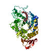
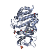
| Entry | Database: PDB / ID: 2dep | ||||||
|---|---|---|---|---|---|---|---|
| Title | Crystal Structure of xylanase B from Clostridium stercorarium F9 | ||||||
 Components Components | Thermostable celloxylanase | ||||||
 Keywords Keywords | HYDROLASE / GLYCOSIDASE / XYLAN DEGRADATION / FAMILY 10 / Structural Genomics / NPPSFA / National Project on Protein Structural and Functional Analyses | ||||||
| Function / homology |  Function and homology information Function and homology informationcellulase / cellulase activity / endo-1,4-beta-xylanase / endo-1,4-beta-xylanase activity / xylan catabolic process / cellulose catabolic process Similarity search - Function | ||||||
| Biological species |  Clostridium stercorarium (bacteria) Clostridium stercorarium (bacteria) | ||||||
| Method |  X-RAY DIFFRACTION / MOLECULAR REPLACEMENT / Resolution: 1.8 Å X-RAY DIFFRACTION / MOLECULAR REPLACEMENT / Resolution: 1.8 Å | ||||||
 Authors Authors | Fushinobu, S. / Nishimoto, M. / Miyanaga, A. / Kitaoka, M. / Hayashi, K. | ||||||
 Citation Citation | Journal: To be Published Title: Molecular anatomy of the alkaliphilic xylanase from Bacillus halodurans C-125 Authors: Nishimoto, M. / Fushinobu, S. / Miyanaga, A. / Kitaoka, M. / Hayashi, K. #1: Journal: ACTA CRYSTALLOGR.,SECT.D / Year: 2004 Title: Crystallization and preliminary X-ray analysis of xylanase B from Clostridium stercorarium Authors: Nishimoto, M. / Fushinobu, S. / Miyanaga, A. / Wakagi, T. / Shoun, H. / Sakka, K. / Ohmiya, K. / Nirasawa, S. / Kitaoka, M. / Hayashi, K. #2: Journal: Biosci.Biotechnol.Biochem. / Year: 2005 Title: The role of conserved arginine residue in loop 4 of glycoside hydrolase family 10 xylanases Authors: Nishimoto, M. / Kitaoka, M. / Fushinobu, S. / Hayashi, K. #3: Journal: J.Biosci.Bioeng. / Year: 2002 Title: Employing chimeric xylanases to identify regions of an alkaline xylanase participating in enzyme activity at basic pH Authors: Nishimoto, M. / Kitaoka, M. / Hayashi, K. | ||||||
| History |
|
- Structure visualization
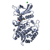
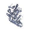
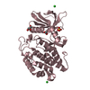
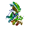
| Structure viewer | Molecule: MolmilJmol/JSmol |
|---|
- Downloads & links
-Download
| PDBx/mmCIF format | 2dep.cif.gz | 163.1 KB | Display | PDBx/mmCIF format |
|---|---|---|---|---|
| PDB format | pdb2dep.ent.gz | 126.9 KB | Display | PDB format |
| PDBx/mmJSON format | 2dep.json.gz | Tree view | PDBx/mmJSON format | |
| Others |  Other downloads Other downloads |
-Validation report
| Arichive directory | https://data.pdbj.org/pub/pdb/validation_reports/de/2depftp://data.pdbj.org/pub/pdb/validation_reports/de/2dep | HTTPS FTP |
|---|
-Related structure data
| Related structure data |  1hizS S: Starting model for refinement |
|---|---|
| Similar structure data |


-Links
 PDBj
PDBj
- Assembly
| Deposited unit | 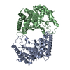
| ||||||||
|---|---|---|---|---|---|---|---|---|---|
| 1 | 
| ||||||||
| 2 | 
| ||||||||
| Unit cell |
|
-Components
| #1: Protein | Mass: 41365.602 Da / Num. of mol.: 2 / Fragment: residues 41-387 Source method: isolated from a genetically manipulated source Source: (gene. exp.) Clostridium stercorarium (bacteria) / Strain: F-9 / Gene: xynB / Plasmid: pET28 / Species (production host): Escherichia coli / Production host: References: UniProt: P40942, cellulase, endo-1,4-beta-xylanase #2: Water | ChemComp-HOH / |  Mass: 18.015 Da / Num. of mol.: 708 / Source method: isolated from a natural source / Formula: H2O Mass: 18.015 Da / Num. of mol.: 708 / Source method: isolated from a natural source / Formula: H2O |
|---|
-Experimental details
-Experiment
| Experiment | Method: X-RAY DIFFRACTION / Number of used crystals: 1 |
|---|
- Sample preparation
| Crystal | Density Matthews: 2.62 Å3/Da / Density % sol: 52.98 % |
|---|---|
| Crystal grow | Temperature: 293 K / Method: vapor diffusion, sitting drop / pH: 6.5 Details: 24% PEG 1500, 20% glycerol, pH 6.5, VAPOR DIFFUSION, SITTING DROP, temperature 293K |
-Data collection
| Diffraction | Mean temperature: 100 K |
|---|---|
| Diffraction source | Source: ROTATING ANODE / Type: RIGAKU ULTRAX 18 / Wavelength: 1.5418 Å |
| Detector | Type: RIGAKU RAXIS IV / Detector: IMAGE PLATE / Date: Jul 23, 2003 / Details: mirrors |
| Radiation | Monochromator: YALE MIRRORS / Protocol: SINGLE WAVELENGTH / Monochromatic (M) / Laue (L): M / Scattering type: x-ray |
| Radiation wavelength | Wavelength: 1.5418 Å / Relative weight: 1 |
| Reflection | Resolution: 1.8→50.14 Å / Num. all: 81152 / Num. obs: 80942 / % possible obs: 99.7 % / Observed criterion σ(F): 0 / Observed criterion σ(I): 3 / Redundancy: 6.1 % / Biso Wilson estimate: 18.8 Å2 / Rmerge(I) obs: 0.062 / Net I/σ(I): 8.8 |
| Reflection shell | Resolution: 1.8→1.86 Å / Redundancy: 5.7 % / Rmerge(I) obs: 0.276 / Mean I/σ(I) obs: 2.5 / Num. unique all: 8015 / % possible all: 99.7 |
- Processing
| Software |
| ||||||||||||||||||||||||||||||||||||||||||||||||||||||||||||||||||||||||||||||||
|---|---|---|---|---|---|---|---|---|---|---|---|---|---|---|---|---|---|---|---|---|---|---|---|---|---|---|---|---|---|---|---|---|---|---|---|---|---|---|---|---|---|---|---|---|---|---|---|---|---|---|---|---|---|---|---|---|---|---|---|---|---|---|---|---|---|---|---|---|---|---|---|---|---|---|---|---|---|---|---|---|---|
| Refinement | Method to determine structure: MOLECULAR REPLACEMENT Starting model: PDB ENTRY 1HIZ Resolution: 1.8→50.14 Å / Rfactor Rfree error: 0.003 / Data cutoff high absF: 2018982.65 / Data cutoff low absF: 0 / Isotropic thermal model: RESTRAINED / Cross valid method: THROUGHOUT / σ(F): 0 / Stereochemistry target values: Engh & Huber
| ||||||||||||||||||||||||||||||||||||||||||||||||||||||||||||||||||||||||||||||||
| Solvent computation | Solvent model: FLAT MODEL / Bsol: 51.5148 Å2 / ksol: 0.388148 e/Å3 | ||||||||||||||||||||||||||||||||||||||||||||||||||||||||||||||||||||||||||||||||
| Displacement parameters | Biso mean: 20.2 Å2
| ||||||||||||||||||||||||||||||||||||||||||||||||||||||||||||||||||||||||||||||||
| Refine analyze |
| ||||||||||||||||||||||||||||||||||||||||||||||||||||||||||||||||||||||||||||||||
| Refinement step | Cycle: LAST / Resolution: 1.8→50.14 Å
| ||||||||||||||||||||||||||||||||||||||||||||||||||||||||||||||||||||||||||||||||
| Refine LS restraints |
| ||||||||||||||||||||||||||||||||||||||||||||||||||||||||||||||||||||||||||||||||
| LS refinement shell | Resolution: 1.8→1.91 Å / Rfactor Rfree error: 0.01 / Total num. of bins used: 6
| ||||||||||||||||||||||||||||||||||||||||||||||||||||||||||||||||||||||||||||||||
| Xplor file |
|
