- PDB-2db7: Crystal structure of hypothetical protein MS0332 -
+
Open data
ID or keywords:
Loading...
-
Basic information
Entry
Database: PDB / ID: 2db7
Title
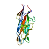
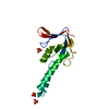
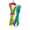
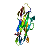
Crystal structure of hypothetical protein MS0332
Components
Hairy/enhancer-of-split related with YRPW motif 1
Keywords
TRANSCRIPTION / STRUCTURAL GENOMICS / UNKNOWN FUNCTION / DNA BINDING PROTEIN / NPPSFA / National Project on Protein Structural and Functional Analyses / RIKEN Structural Genomics/Proteomics Initiative / RSGI
Function / homology
Function and homology information
umbilical cord morphogenesis / regulation of vasculogenesis / atrioventricular valve formation / dorsal aorta morphogenesis / arterial endothelial cell differentiation / negative regulation of smooth muscle cell differentiation / cardiac ventricle morphogenesis / cardiac epithelial to mesenchymal transition / cardiac conduction system development / pulmonary valve morphogenesis ...umbilical cord morphogenesis / regulation of vasculogenesis / atrioventricular valve formation / dorsal aorta morphogenesis / arterial endothelial cell differentiation / negative regulation of smooth muscle cell differentiation / cardiac ventricle morphogenesis / cardiac epithelial to mesenchymal transition / cardiac conduction system development / pulmonary valve morphogenesis / negative regulation of biomineral tissue development / heart trabecula formation / circulatory system development / aortic valve morphogenesis / Cardiogenesis / NOTCH4 Intracellular Domain Regulates Transcription / labyrinthine layer blood vessel development / NOTCH3 Intracellular Domain Regulates Transcription / endocardial cushion morphogenesis / ventricular septum morphogenesis / negative regulation of neuron differentiation / cardiac septum morphogenesis / RUNX2 regulates osteoblast differentiation / negative regulation of Notch signaling pathway / regulation of neurogenesis / cis-regulatory region sequence-specific DNA binding / Notch signaling pathway / NOTCH1 Intracellular Domain Regulates Transcription / DNA-binding transcription repressor activity, RNA polymerase II-specific / Constitutive Signaling by NOTCH1 PEST Domain Mutants / Constitutive Signaling by NOTCH1 HD+PEST Domain Mutants / sequence-specific double-stranded DNA binding / angiogenesis / RNA polymerase II-specific DNA-binding transcription factor binding / DNA-binding transcription factor activity, RNA polymerase II-specific / protein dimerization activity / RNA polymerase II cis-regulatory region sequence-specific DNA binding / DNA-binding transcription factor activity / negative regulation of DNA-templated transcription / chromatin / negative regulation of transcription by RNA polymerase II / positive regulation of transcription by RNA polymerase II / nucleoplasm / nucleus / cytoplasm Similarity search - Function
Single alpha-helices involved in coiled-coils or other helix-helix interfaces - #980 / Orange domain / : / Hairy Orange / Orange domain profile. / Orange domain / Helix-loop-helix DNA-binding domain / Single alpha-helices involved in coiled-coils or other helix-helix interfaces / helix loop helix domain / Myc-type, basic helix-loop-helix (bHLH) domain ...Single alpha-helices involved in coiled-coils or other helix-helix interfaces - #980 / Orange domain / : / Hairy Orange / Orange domain profile. / Orange domain / Helix-loop-helix DNA-binding domain / Single alpha-helices involved in coiled-coils or other helix-helix interfaces / helix loop helix domain / Myc-type, basic helix-loop-helix (bHLH) domain / Myc-type, basic helix-loop-helix (bHLH) domain profile. / Helix-loop-helix DNA-binding domain superfamily / Helix non-globular / Special Similarity search - Domain/homology
In the structure databanks used in Yorodumi, some data are registered as the other names, "COVID-19 virus" and "2019-nCoV". Here are the details of the virus and the list of structure data.
Jan 31, 2019. EMDB accession codes are about to change! (news from PDBe EMDB page)
EMDB accession codes are about to change! (news from PDBe EMDB page)
The allocation of 4 digits for EMDB accession codes will soon come to an end. Whilst these codes will remain in use, new EMDB accession codes will include an additional digit and will expand incrementally as the available range of codes is exhausted. The current 4-digit format prefixed with “EMD-” (i.e. EMD-XXXX) will advance to a 5-digit format (i.e. EMD-XXXXX), and so on. It is currently estimated that the 4-digit codes will be depleted around Spring 2019, at which point the 5-digit format will come into force.
The EM Navigator/Yorodumi systems omit the EMD- prefix.
Related info.:Q: What is EMD? / ID/Accession-code notation in Yorodumi/EM Navigator
Yorodumi is a browser for structure data from EMDB, PDB, SASBDB, etc.
This page is also the successor to EM Navigator detail page, and also detail information page/front-end page for Omokage search.
The word "yorodu" (or yorozu) is an old Japanese word meaning "ten thousand". "mi" (miru) is to see.
Related info.:EMDB / PDB / SASBDB / Comparison of 3 databanks / Yorodumi Search / Aug 31, 2016. New EM Navigator & Yorodumi / Yorodumi Papers / Jmol/JSmol / Function and homology information / Changes in new EM Navigator and Yorodumi
 Movie
Movie Controller


 Open data
 Basic information
 Components
 Keywords
 Function and homology information
 Homo sapiens (human)
 X-RAY DIFFRACTION /
  Authors
 Citation
 Structure visualization
 Downloads & links
 Other downloads
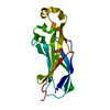


 PDBj
PDBj


 Assembly
 Mass: 18.015 Da / Num. of mol.: 97 / Source method: isolated from a natural source / Formula: H2O
 Sample preparation
 / Beamline: BL26B1 / Wavelength: 0.9789, 0.9791, 0.9650
 Processing