 Movie
Movie Controller

[English] 日本語
 Yorodumi
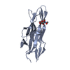
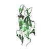
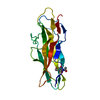
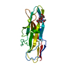
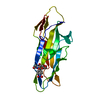
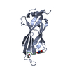
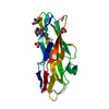
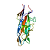
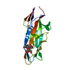
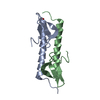
- PDB-1zk5: Escherichia coli F17fG lectin domain complex with N-acetylglucosamine -
+ Open data

- Basic information
| Entry | Database: PDB / ID: 1zk5 | ||||||
|---|---|---|---|---|---|---|---|
| Title | Escherichia coli F17fG lectin domain complex with N-acetylglucosamine | ||||||
 Components Components | F17G adhesin subunit | ||||||
 Keywords Keywords | CELL ADHESION / lectin / beta sandwich / protein-structure complex / immunoglobulin-like fold | ||||||
| Function / homology |  Function and homology information Function and homology informationadhesion of symbiont to host / cell adhesion involved in single-species biofilm formation / pilus / carbohydrate binding Similarity search - Function | ||||||
| Biological species |  | ||||||
| Method |  X-RAY DIFFRACTION / SYNCHROTRON / MOLECULAR REPLACEMENT / Resolution: 1.4 Å X-RAY DIFFRACTION / SYNCHROTRON / MOLECULAR REPLACEMENT / Resolution: 1.4 Å | ||||||
 Authors Authors | Buts, L. / Wellens, A. / Van Molle, I. / De Genst, E. / Wyns, L. / Loris, R. / Lahmann, M. / Oscarson, S. / De Greve, H. / Bouckaert, J. | ||||||
 Citation Citation | Journal: Acta Crystallogr.,Sect.D / Year: 2005 Title: Impact of natural variation in bacterial F17G adhesins on crystallization behaviour. Authors: Buts, L. / Wellens, A. / Van Molle, I. / Wyns, L. / Loris, R. / Lahmann, M. / Oscarson, S. / De Greve, H. / Bouckaert, J. | ||||||
| History |
|
- Structure visualization
| Structure viewer | Molecule: MolmilJmol/JSmol |
|---|
- Downloads & links
-Download
| PDBx/mmCIF format | 1zk5.cif.gz | 51.5 KB | Display | PDBx/mmCIF format |
|---|---|---|---|---|
| PDB format | pdb1zk5.ent.gz | 35.5 KB | Display | PDB format |
| PDBx/mmJSON format | 1zk5.json.gz | Tree view | PDBx/mmJSON format | |
| Others |  Other downloads Other downloads |
-Validation report
| Arichive directory | https://data.pdbj.org/pub/pdb/validation_reports/zk/1zk5ftp://data.pdbj.org/pub/pdb/validation_reports/zk/1zk5 | HTTPS FTP |
|---|
-Related structure data
| Related structure data |  1zplC  2bs7C  2bs8C  2bsbC 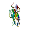 2bscC 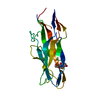 1o9vS S: Starting model for refinement C: citing same article ( |
|---|---|
| Similar structure data |
-Links
 PDBj
PDBj- Assembly
| Deposited unit | 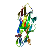
| ||||||||
|---|---|---|---|---|---|---|---|---|---|
| 1 |
| ||||||||
| Unit cell |
| ||||||||
| Components on special symmetry positions |
|
-Components
| #1: Protein | Mass: 18859.789 Da / Num. of mol.: 1 / Fragment: residues 23-198 Source method: isolated from a genetically manipulated source Source: (gene. exp.) |
|---|---|
| #2: Sugar | ChemComp-NAG /   Type: D-saccharide, beta linking / Mass: 221.208 Da / Num. of mol.: 1 Type: D-saccharide, beta linking / Mass: 221.208 Da / Num. of mol.: 1Source method: isolated from a genetically manipulated source Formula: C8H15NO6 |
| #3: Water | ChemComp-HOH /  Mass: 18.015 Da / Num. of mol.: 229 / Source method: isolated from a natural source / Formula: H2O Mass: 18.015 Da / Num. of mol.: 229 / Source method: isolated from a natural source / Formula: H2O |
| Has protein modification | Y |
-Experimental details
-Experiment
| Experiment | Method: X-RAY DIFFRACTION / Number of used crystals: 1 |
|---|
- Sample preparation
| Crystal | Density Matthews: 2.17 Å3/Da / Density % sol: 43.35 % |
|---|---|
| Crystal grow | Temperature: 298 K / Method: vapor diffusion, hanging drop / pH: 5.6 Details: 20% (v/v) isopropanol, 20% PEG 4000, 100 mM Na citrate, pH 5.6, VAPOR DIFFUSION, HANGING DROP, temperature 298K |
-Data collection
| Diffraction | Mean temperature: 100 K |
|---|---|
| Diffraction source | Source: SYNCHROTRON / Site: EMBL/DESY, HAMBURG  / Beamline: X11 / Wavelength: 0.811 Å / Beamline: X11 / Wavelength: 0.811 Å |
| Detector | Type: MARRESEARCH / Detector: CCD / Date: Nov 28, 2004 |
| Radiation | Monochromator: DESY X11 / Protocol: SINGLE WAVELENGTH / Monochromatic (M) / Laue (L): M / Scattering type: x-ray |
| Radiation wavelength | Wavelength: 0.811 Å / Relative weight: 1 |
| Reflection | Resolution: 1.38→50 Å / Num. all: 33091 / Num. obs: 32992 / % possible obs: 99.7 % / Observed criterion σ(F): 0 / Observed criterion σ(I): 0 / Biso Wilson estimate: 11.3 Å2 / Rmerge(I) obs: 0.048 / Net I/σ(I): 15.7 |
| Reflection shell | Resolution: 1.38→1.43 Å / Rmerge(I) obs: 0.242 / % possible all: 99 |
- Processing
| Software |
| ||||||||||||||||||||
|---|---|---|---|---|---|---|---|---|---|---|---|---|---|---|---|---|---|---|---|---|---|
| Refinement | Method to determine structure: MOLECULAR REPLACEMENT Starting model: PDB entry 1O9V Resolution: 1.4→50 Å / Cross valid method: THROUGHOUT / σ(F): 0 / Stereochemistry target values: Engh & Huber
| ||||||||||||||||||||
| Refinement step | Cycle: LAST / Resolution: 1.4→50 Å
| ||||||||||||||||||||
| LS refinement shell | Resolution: 1.4→1.47 Å / Rfactor Rfree error: 0.013
|
