 ムービー
ムービー コントローラー


+ データを開く

- 基本情報
基本情報
| 登録情報 | データベース: PDB / ID: 2d4c | ||||||
|---|---|---|---|---|---|---|---|
| タイトル | Crystal structure of the endophilin BAR domain mutant | ||||||
 要素 要素 | SH3-containing GRB2-like protein 2 | ||||||
 キーワード キーワード | TRANSFERASE / BAR domain | ||||||
| 機能・相同性 |  機能・相同性情報 機能・相同性情報postsynaptic actin cytoskeleton organization / negative regulation of blood-brain barrier permeability / dendrite extension / regulation of receptor internalization / photoreceptor ribbon synapse / Retrograde neurotrophin signalling / Lysosome Vesicle Biogenesis / Golgi Associated Vesicle Biogenesis / negative regulation of epidermal growth factor receptor signaling pathway / Recycling pathway of L1 ...postsynaptic actin cytoskeleton organization / negative regulation of blood-brain barrier permeability / dendrite extension / regulation of receptor internalization / photoreceptor ribbon synapse / Retrograde neurotrophin signalling / Lysosome Vesicle Biogenesis / Golgi Associated Vesicle Biogenesis / negative regulation of epidermal growth factor receptor signaling pathway / Recycling pathway of L1 / synaptic vesicle endocytosis / presynaptic cytosol / negative regulation of MAPK cascade / cellular response to brain-derived neurotrophic factor stimulus / MHC class II antigen presentation / InlB-mediated entry of Listeria monocytogenes into host cell / central nervous system development / cell projection / EGFR downregulation / clathrin-coated endocytic vesicle membrane / Negative regulation of MET activity / neuron projection development / Cargo recognition for clathrin-mediated endocytosis / presynapse / Clathrin-mediated endocytosis / early endosome / postsynapse / Golgi membrane / negative regulation of gene expression / lipid binding / perinuclear region of cytoplasm / glutamatergic synapse / signal transduction / identical protein binding / plasma membrane / cytoplasm / cytosol 類似検索 - 分子機能 | ||||||
| 生物種 |  Homo sapiens (ヒト) Homo sapiens (ヒト) | ||||||
| 手法 |  X線回折 / シンクロトロン / 分子置換 / 解像度: 2.4 Å X線回折 / シンクロトロン / 分子置換 / 解像度: 2.4 Å | ||||||
 データ登録者 データ登録者 | Masuda, M. / Takeda, S. | ||||||
 引用 引用 | ジャーナル: Embo J. / 年: 2006 タイトル: Endophilin BAR domain drives membrane curvature by two newly identified structure-based mechanisms 著者: Masuda, M. / Takeda, S. / Sone, M. / Ohki, T. / Mori, H. / Kamioka, Y. / Mochizuki, N. | ||||||
| 履歴 |
|
- 構造の表示
構造の表示
| 構造ビューア | 分子: MolmilJmol/JSmol |
|---|
- ダウンロードとリンク
-ダウンロード
| PDBx/mmCIF形式 | 2d4c.cif.gz | 190.4 KB | 表示 | PDBx/mmCIF形式 |
|---|---|---|---|---|
| PDB形式 | pdb2d4c.ent.gz | 152.7 KB | 表示 | PDB形式 |
| PDBx/mmJSON形式 | 2d4c.json.gz | ツリー表示 | PDBx/mmJSON形式 | |
| その他 |  その他のダウンロード その他のダウンロード |
-検証レポート
| 文書・要旨 | 2d4c_validation.pdf.gz | 461.6 KB | 表示 | wwPDB検証レポート |
|---|---|---|---|---|
| 文書・詳細版 | 2d4c_full_validation.pdf.gz | 481.8 KB | 表示 | |
| XML形式データ | 2d4c_validation.xml.gz | 35.2 KB | 表示 | |
| CIF形式データ | 2d4c_validation.cif.gz | 48.6 KB | 表示 | |
| アーカイブディレクトリ | https://data.pdbj.org/pub/pdb/validation_reports/d4/2d4cftp://data.pdbj.org/pub/pdb/validation_reports/d4/2d4c | HTTPS FTP |
-関連構造データ


-リンク
 PDBj
PDBj


- 集合体
集合体
| 登録構造単位 | 
| ||||||||
|---|---|---|---|---|---|---|---|---|---|
| 1 | 
| ||||||||
| 2 | 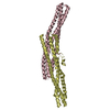
| ||||||||
| 単位格子 |
|
-要素
| #1: タンパク質 | 分子量: 29056.242 Da / 分子数: 4 / 断片: endophilin A1 BAR domain / 由来タイプ: 組換発現 / 由来: (組換発現) Homo sapiens (ヒト) / プラスミド: pGEX6p3 / 発現宿主:  参照: UniProt: Q99962, 転移酵素; アシル基を移すもの; アミノアシル基以外のアシル基を移すもの #2: 化合物 | ChemComp-CA / |   分子量: 40.078 Da / 分子数: 1 / 由来タイプ: 合成 / 式: Ca 分子量: 40.078 Da / 分子数: 1 / 由来タイプ: 合成 / 式: Ca#3: 水 | ChemComp-HOH / |  分子量: 18.015 Da / 分子数: 197 / 由来タイプ: 天然 / 式: H2O 分子量: 18.015 Da / 分子数: 197 / 由来タイプ: 天然 / 式: H2O |
|---|
-実験情報
-実験
| 実験 | 手法: X線回折 / 使用した結晶の数: 1 |
|---|
- 試料調製
試料調製
| 結晶 | マシュー密度: 2.18 Å3/Da / 溶媒含有率: 43.55 % |
|---|---|
| 結晶化 | 温度: 293 K / 手法: 蒸気拡散法, シッティングドロップ法 / pH: 7.2 詳細: 100mM HEPES, 200mM calcium acetate, 10mM DTT, 20% PEG3350, pH 7.2, VAPOR DIFFUSION, SITTING DROP, temperature 293K |
-データ収集
| 回折 | 平均測定温度: 90 K |
|---|---|
| 放射光源 | 由来: シンクロトロン / サイト: SPring-8  / ビームライン: BL38B1 / 波長: 1 Å / ビームライン: BL38B1 / 波長: 1 Å |
| 検出器 | タイプ: RIGAKU / 検出器: IMAGE PLATE / 日付: 2005年6月6日 |
| 放射 | プロトコル: SINGLE WAVELENGTH / 単色(M)・ラウエ(L): M / 散乱光タイプ: x-ray |
| 放射波長 | 波長: 1 Å / 相対比: 1 |
| 反射 | 解像度: 2.4→50 Å / Num. obs: 38763 / % possible obs: 99.6 % / Observed criterion σ(F): 0 / Observed criterion σ(I): 0 / 冗長度: 3.8 % / Rmerge(I) obs: 0.098 / Net I/σ(I): 11 |
| 反射 シェル | 解像度: 2.4→2.49 Å / 冗長度: 3.1 % / Rmerge(I) obs: 0.323 / Num. unique all: 3673 / % possible all: 93.5 |
- 解析
解析
| ソフトウェア |
| |||||||||||||||||||||||||
|---|---|---|---|---|---|---|---|---|---|---|---|---|---|---|---|---|---|---|---|---|---|---|---|---|---|---|
| 精密化 | 構造決定の手法: 分子置換 開始モデル: PDB entry 1X03 解像度: 2.4→50 Å / 交差検証法: THROUGHOUT / σ(F): 0 / 立体化学のターゲット値: Engh & Huber
| |||||||||||||||||||||||||
| Refine analyze |
| |||||||||||||||||||||||||
| 精密化ステップ | サイクル: LAST / 解像度: 2.4→50 Å
| |||||||||||||||||||||||||
| 拘束条件 |
| |||||||||||||||||||||||||
| LS精密化 シェル | 解像度: 2.4→2.49 Å
|
