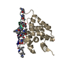
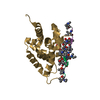
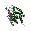
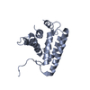
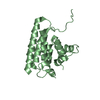
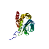
- PDB-2cxl: RUN domain of Rap2 interacting protein x, crystallized in I422 sp... -
+
Open data
ID or keywords:
Loading...
-
Basic information
Entry
Database: PDB / ID: 2cxl
Title
RUN domain of Rap2 interacting protein x, crystallized in I422 space group
Components
rap2 interacting protein x
Keywords
PROTEIN BINDING / RUN domain / helix bundle / Structural Genomics / NPPSFA / National Project on Protein Structural and Functional Analyses / RIKEN Structural Genomics/Proteomics Initiative / RSGI
Function / homology
Function and homology information
positive regulation of retrograde axon cargo transport / endolysosome / negative regulation of axonogenesis / positive regulation of intracellular protein transport / positive regulation of axonogenesis / regulation of axonogenesis / regulation of establishment of cell polarity / dynactin binding / positive regulation of axon extension / actin filament organization ...positive regulation of retrograde axon cargo transport / endolysosome / negative regulation of axonogenesis / positive regulation of intracellular protein transport / positive regulation of axonogenesis / regulation of axonogenesis / regulation of establishment of cell polarity / dynactin binding / positive regulation of axon extension / actin filament organization / filopodium / nervous system development / lamellipodium / growth cone / perikaryon / cell differentiation / positive regulation of cell migration / axon / neuronal cell body / dendrite / cytosol / cytoplasm Similarity search - Function
: / : / RUN domain / RUN / RUN domain / RUN domain superfamily / RUN domain / RUN domain profile. / Methane Monooxygenase Hydroxylase; Chain G, domain 1 / Up-down Bundle / Mainly Alpha Similarity search - Domain/homology
In the structure databanks used in Yorodumi, some data are registered as the other names, "COVID-19 virus" and "2019-nCoV". Here are the details of the virus and the list of structure data.
Jan 31, 2019. EMDB accession codes are about to change! (news from PDBe EMDB page)
EMDB accession codes are about to change! (news from PDBe EMDB page)
The allocation of 4 digits for EMDB accession codes will soon come to an end. Whilst these codes will remain in use, new EMDB accession codes will include an additional digit and will expand incrementally as the available range of codes is exhausted. The current 4-digit format prefixed with “EMD-” (i.e. EMD-XXXX) will advance to a 5-digit format (i.e. EMD-XXXXX), and so on. It is currently estimated that the 4-digit codes will be depleted around Spring 2019, at which point the 5-digit format will come into force.
The EM Navigator/Yorodumi systems omit the EMD- prefix.
Related info.:Q: What is EMD? / ID/Accession-code notation in Yorodumi/EM Navigator
Yorodumi is a browser for structure data from EMDB, PDB, SASBDB, etc.
This page is also the successor to EM Navigator detail page, and also detail information page/front-end page for Omokage search.
The word "yorodu" (or yorozu) is an old Japanese word meaning "ten thousand". "mi" (miru) is to see.
Related info.:EMDB / PDB / SASBDB / Comparison of 3 databanks / Yorodumi Search / Aug 31, 2016. New EM Navigator & Yorodumi / Yorodumi Papers / Jmol/JSmol / Function and homology information / Changes in new EM Navigator and Yorodumi
 Movie
Movie Controller

 Yorodumi
 Open data
 Basic information
 Components
 Keywords
 Function and homology information
 X-RAY DIFFRACTION /
  Authors
 Citation
 Structure visualization
 Downloads & links
 Other downloads


 PDBj
PDBj Assembly
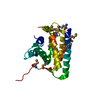
 Sample preparation
 / Beamline: BL26B1 / Wavelength: 0.97888, 0.97951, 0.97600, 0.98300
 Processing