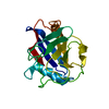
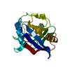
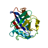
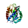
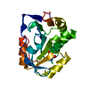
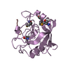
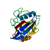
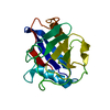
Entry Database : PDB / ID : 2ck1Title The structure of oxidised cyclophilin A from s. mansoni PEPTIDYL-PROLYL CIS-TRANS ISOMERASE E Keywords / / / / / / / Function / homology Function Domain/homology Component
/ / / / / / / / / / / / / / / / / / / / / / / / / / / / / Biological species SCHISTOSOMA MANSONI (invertebrata)Method / / / Resolution : 1.8 Å Authors Gourlay, L.J. / Angelucci, F. / Bellelli, A. / Boumis, G. / Miele, A.E. / Brunori, M. Journal : J.Biol.Chem. / Year : 2007Title : The Three-Dimensional Structure of Two Redox States of Cyclophilin-A from Schistosoma Mansoni: Evidence for Redox- Regulation of Peptidyl-Prolyl Cis-Trans Isomerase Activity.Authors : Gourlay, L.J. / Angelucci, F. / Baiocco, P. / Boumis, G. / Brunori, M. / Bellelli, A. / Miele, A.E. History Deposition Apr 10, 2006 Deposition site / Processing site Revision 1.0 May 15, 2007 Provider / Type Revision 1.1 May 8, 2011 Group Revision 1.2 Jul 13, 2011 Group Revision 1.3 Dec 13, 2023 Group Data collection / Database references ... Data collection / Database references / Other / Refinement description Category chem_comp_atom / chem_comp_bond ... chem_comp_atom / chem_comp_bond / database_2 / pdbx_database_status / pdbx_initial_refinement_model Item / _database_2.pdbx_database_accession / _pdbx_database_status.status_code_sfRevision 1.4 Nov 6, 2024 Group / Category / pdbx_modification_feature / Item
Show all Show less
 Movie
Movie Controller


 Open data
 Basic information
 Components
 Keywords
 Function and homology information
 X-RAY DIFFRACTION /
  Authors
 Citation
 Structure visualization
 Downloads & links
 Other downloads
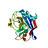


 PDBj
PDBj

 Assembly
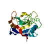


 Mass: 59.044 Da / Num. of mol.: 1 / Source method: obtained synthetically / Formula: C2H3O2
 Mass: 18.015 Da / Num. of mol.: 101 / Source method: isolated from a natural source / Formula: H2O
 Sample preparation
 / Beamline: 5.2R / Wavelength: 1.2
  Processing