 Movie
Movie Controller


+ Open data

- Basic information
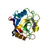
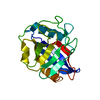
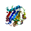
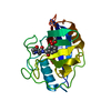
| Entry | Database: PDB / ID: 3k0r | ||||||
|---|---|---|---|---|---|---|---|
| Title | Cryogenic structure of CypA mutant Arg55Lys | ||||||
 Components Components | Cyclophilin A | ||||||
 Keywords Keywords | ISOMERASE / proline isomerase / Acetylation / Cyclosporin / Cytoplasm / Host-virus interaction / Isopeptide bond / Phosphoprotein / Rotamase / Ubl conjugation | ||||||
| Function / homology |  Function and homology information Function and homology informationnegative regulation of protein K48-linked ubiquitination / regulation of apoptotic signaling pathway / cell adhesion molecule production / lipid droplet organization / negative regulation of viral life cycle / heparan sulfate binding / regulation of viral genome replication / virion binding / negative regulation of stress-activated MAPK cascade / activation of protein kinase B activity ...negative regulation of protein K48-linked ubiquitination / regulation of apoptotic signaling pathway / cell adhesion molecule production / lipid droplet organization / negative regulation of viral life cycle / heparan sulfate binding / regulation of viral genome replication / virion binding / negative regulation of stress-activated MAPK cascade / activation of protein kinase B activity / endothelial cell activation / leukocyte chemotaxis / Basigin interactions / protein peptidyl-prolyl isomerization / cyclosporin A binding / Minus-strand DNA synthesis / Plus-strand DNA synthesis / Uncoating of the HIV Virion / Early Phase of HIV Life Cycle / Integration of provirus / APOBEC3G mediated resistance to HIV-1 infection / negative regulation of protein phosphorylation / viral release from host cell / Binding and entry of HIV virion / Calcineurin activates NFAT / negative regulation of oxidative stress-induced intrinsic apoptotic signaling pathway / negative regulation of protein kinase activity / positive regulation of viral genome replication / neutrophil chemotaxis / : / Gene and protein expression by JAK-STAT signaling after Interleukin-12 stimulation / positive regulation of protein secretion / peptidylprolyl isomerase / peptidyl-prolyl cis-trans isomerase activity / Assembly Of The HIV Virion / platelet activation / Budding and maturation of HIV virion / positive regulation of protein phosphorylation / platelet aggregation / integrin binding / neuron differentiation / SARS-CoV-1 activates/modulates innate immune responses / : / Platelet degranulation / cellular response to oxidative stress / protein folding / secretory granule lumen / vesicle / ficolin-1-rich granule lumen / positive regulation of MAPK cascade / focal adhesion / apoptotic process / Neutrophil degranulation / protein-containing complex / : / RNA binding / extracellular exosome / extracellular region / membrane / nucleus / cytosol / cytoplasm Similarity search - Function | ||||||
| Biological species |  Homo sapiens (human) Homo sapiens (human) | ||||||
| Method |  X-RAY DIFFRACTION / SYNCHROTRON / MOLECULAR REPLACEMENT / Resolution: 2.424 Å X-RAY DIFFRACTION / SYNCHROTRON / MOLECULAR REPLACEMENT / Resolution: 2.424 Å | ||||||
 Authors Authors | Fraser, J.S. / Alber, T. | ||||||
 Citation Citation | Journal: Nature / Year: 2009 Title: Hidden alternative structures of proline isomerase essential for catalysis. Authors: Fraser, J.S. / Clarkson, M.W. / Degnan, S.C. / Erion, R. / Kern, D. / Alber, T. | ||||||
| History |
|
- Structure visualization
| Structure viewer | Molecule: MolmilJmol/JSmol |
|---|
- Downloads & links
-Download
| PDBx/mmCIF format | 3k0r.cif.gz | 71.3 KB | Display | PDBx/mmCIF format |
|---|---|---|---|---|
| PDB format | pdb3k0r.ent.gz | 53.9 KB | Display | PDB format |
| PDBx/mmJSON format | 3k0r.json.gz | Tree view | PDBx/mmJSON format | |
| Others |  Other downloads Other downloads |
-Validation report
| Arichive directory | https://data.pdbj.org/pub/pdb/validation_reports/k0/3k0rftp://data.pdbj.org/pub/pdb/validation_reports/k0/3k0r | HTTPS FTP |
|---|
-Related structure data
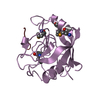
| Related structure data |  3k0mC 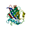 3k0nC  3k0oC  3k0pC  3k0qC  2cplS C: citing same article ( S: Starting model for refinement |
|---|---|
| Similar structure data |


-Links
 PDBj
PDBj


- Assembly
| Deposited unit | 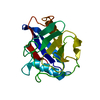
| ||||||||
|---|---|---|---|---|---|---|---|---|---|
| 1 |
| ||||||||
| Unit cell |
|
-Components
| #1: Protein | Mass: 18008.488 Da / Num. of mol.: 1 / Mutation: R55K Source method: isolated from a genetically manipulated source Source: (gene. exp.) Homo sapiens (human) / Gene: PPIA, CYPA / Production host:  |
|---|---|
| #2: Water | ChemComp-HOH /  Mass: 18.015 Da / Num. of mol.: 59 / Source method: isolated from a natural source / Formula: H2O Mass: 18.015 Da / Num. of mol.: 59 / Source method: isolated from a natural source / Formula: H2O |
-Experimental details
-Experiment
| Experiment | Method: X-RAY DIFFRACTION / Number of used crystals: 1 |
|---|
- Sample preparation
| Crystal | Density Matthews: 3.09 Å3/Da / Density % sol: 60.22 % |
|---|---|
| Crystal grow | Temperature: 291 K / Method: vapor diffusion, hanging drop / pH: 7 Details: 1.8 M DL-malic acid, pH 7, VAPOR DIFFUSION, HANGING DROP, temperature 291K |
-Data collection
| Diffraction | Mean temperature: 100 K |
|---|---|
| Diffraction source | Source: SYNCHROTRON / Site: ALS  / Beamline: 8.3.1 / Wavelength: 1.1158 Å / Beamline: 8.3.1 / Wavelength: 1.1158 Å |
| Detector | Type: ADSC QUANTUM 315 / Detector: CCD / Date: Nov 5, 2008 |
| Radiation | Monochromator: SI 111 / Protocol: SINGLE WAVELENGTH / Monochromatic (M) / Laue (L): M / Scattering type: x-ray |
| Radiation wavelength | Wavelength: 1.1158 Å / Relative weight: 1 |
| Reflection | Resolution: 2.42→50 Å / Num. all: 8900 / Num. obs: 8900 / % possible obs: 100 % / Observed criterion σ(F): 0 / Observed criterion σ(I): 0 / Redundancy: 9.8 % / Rsym value: 0.131 / Net I/σ(I): 16.4 |
| Reflection shell | Resolution: 2.42→2.51 Å / Redundancy: 9.8 % / Mean I/σ(I) obs: 3.9 / Num. unique all: 853 / Rsym value: 0.572 / % possible all: 100 |
- Processing
| Software |
| |||||||||||||||||||||||||||||||||||||||||||||||||
|---|---|---|---|---|---|---|---|---|---|---|---|---|---|---|---|---|---|---|---|---|---|---|---|---|---|---|---|---|---|---|---|---|---|---|---|---|---|---|---|---|---|---|---|---|---|---|---|---|---|---|
| Refinement | Method to determine structure: MOLECULAR REPLACEMENT Starting model: PDB entry 2cpl Resolution: 2.424→47.803 Å / SU ML: 0.28 / Cross valid method: THROUGHOUT / σ(F): 0.17 / σ(I): 0 / Phase error: 22.04 / Stereochemistry target values: ML
| |||||||||||||||||||||||||||||||||||||||||||||||||
| Solvent computation | Shrinkage radii: 0.9 Å / VDW probe radii: 1.11 Å / Solvent model: FLAT BULK SOLVENT MODEL / Bsol: 31.657 Å2 / ksol: 0.436 e/Å3 | |||||||||||||||||||||||||||||||||||||||||||||||||
| Displacement parameters |
| |||||||||||||||||||||||||||||||||||||||||||||||||
| Refinement step | Cycle: LAST / Resolution: 2.424→47.803 Å
| |||||||||||||||||||||||||||||||||||||||||||||||||
| Refine LS restraints |
| |||||||||||||||||||||||||||||||||||||||||||||||||
| LS refinement shell |
|
