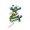
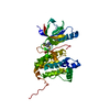
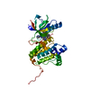
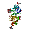
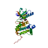
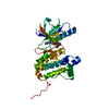
[English] 日本語
Yorodumi
- PDB-2c9a: Crystal structure of the MAM-Ig module of receptor protein tyrosi... -

+
Open data


ID or keywords:

Loading...

-
Basic information

Entry
Database: PDB / ID: 2c9a
TitleCrystal structure of the MAM-Ig module of receptor protein tyrosine phosphatase mu
ComponentsRECEPTOR-TYPE TYROSINE-PROTEIN PHOSPHATASE MU
KeywordsHYDROLASE / GLYCOPROTEIN / IMMUNOGLOBULIN DOMAIN / RECEPTOR
Function / homology
Function and homology information


retina layer formation / transmembrane receptor protein tyrosine phosphatase activity / negative regulation of endothelial cell migration / retinal ganglion cell axon guidance / negative regulation of endothelial cell proliferation / phosphatase activity / homophilic cell adhesion via plasma membrane adhesion molecules / negative regulation of angiogenesis / protein dephosphorylation / protein-tyrosine-phosphatase ...retina layer formation / transmembrane receptor protein tyrosine phosphatase activity / negative regulation of endothelial cell migration / retinal ganglion cell axon guidance / negative regulation of endothelial cell proliferation / phosphatase activity / homophilic cell adhesion via plasma membrane adhesion molecules / negative regulation of angiogenesis / protein dephosphorylation / protein-tyrosine-phosphatase / protein tyrosine phosphatase activity / adherens junction / neuron projection development / cell-cell junction / lamellipodium / response to xenobiotic stimulus / cadherin binding / perinuclear region of cytoplasm / signal transduction / identical protein binding / plasma membrane / cytoplasm
Similarity search - Function
Receptor-type tyrosine-protein phosphatase mu, PTPase domain, repeat 1 / MAM domain signature. / Domain in meprin, A5, receptor protein tyrosine phosphatase mu (and others) / MAM domain, meprin/A5/mu / MAM domain / MAM domain profile. / Immunoglobulin / Immunoglobulin domain / Protein tyrosine phosphatase, catalytic domain / PTP type protein phosphatase domain profile. ...Receptor-type tyrosine-protein phosphatase mu, PTPase domain, repeat 1 / MAM domain signature. / Domain in meprin, A5, receptor protein tyrosine phosphatase mu (and others) / MAM domain, meprin/A5/mu / MAM domain / MAM domain profile. / Immunoglobulin / Immunoglobulin domain / Protein tyrosine phosphatase, catalytic domain / PTP type protein phosphatase domain profile. / Protein-tyrosine phosphatase / Tyrosine-specific protein phosphatase, PTPase domain / Protein-tyrosine phosphatase, catalytic / Protein tyrosine phosphatase, catalytic domain motif / Tyrosine specific protein phosphatases active site. / Protein-tyrosine phosphatase, active site / Tyrosine-specific protein phosphatases domain / Tyrosine specific protein phosphatases domain profile. / Protein-tyrosine phosphatase-like / Jelly Rolls - #200 / Fibronectin type III domain / Fibronectin type 3 domain / Fibronectin type-III domain profile. / Fibronectin type III / Fibronectin type III superfamily / Concanavalin A-like lectin/glucanase domain superfamily / Immunoglobulin subtype / Immunoglobulin / Jelly Rolls / Ig-like domain profile. / Immunoglobulin-like domain / Immunoglobulin-like domain superfamily / Immunoglobulins / Immunoglobulin-like fold / Immunoglobulin-like / Sandwich / Mainly Beta
Similarity search - Domain/homology
Receptor-type tyrosine-protein phosphatase mu
Similarity search - Component
Biological speciesHOMO SAPIENS (human)
MethodX-RAY DIFFRACTION / SYNCHROTRON / MAD / Resolution: 2.7 Å
AuthorsAricescu, A.R. / Hon, W.C. / Siebold, C. / Lu, W. / Van Der Merwe, P.A. / Jones, E.Y.
CitationJournal: Embo J. / Year: 2006
Title: Molecular Analysis of Receptor Protein Tyrosine Phosphatase Mu-Mediated Cell Adhesion.
Authors: Aricescu, A.R. / Hon, W.C. / Siebold, C. / Lu, W. / Van Der Merwe, P.A. / Jones, E.Y.
History
DepositionDec 9, 2005Deposition site: PDBE / Processing site: PDBE
Revision 1.0Jan 19, 2006Provider: repository / Type: Initial release
Revision 1.1Jul 13, 2011Group: Advisory / Version format compliance
Revision 2.0Sep 11, 2019Group: Advisory / Atomic model ...Advisory / Atomic model / Data collection / Derived calculations / Experimental preparation / Non-polymer description / Structure summary
Category: atom_site / chem_comp ...atom_site / chem_comp / database_PDB_caveat / entity / exptl_crystal_grow / pdbx_entity_nonpoly / pdbx_nonpoly_scheme / pdbx_validate_chiral / struct_asym / struct_conn / struct_site_gen
Item: _atom_site.B_iso_or_equiv / _atom_site.Cartn_x ..._atom_site.B_iso_or_equiv / _atom_site.Cartn_x / _atom_site.Cartn_y / _atom_site.Cartn_z / _atom_site.auth_atom_id / _atom_site.auth_comp_id / _atom_site.label_atom_id / _atom_site.label_comp_id / _atom_site.label_entity_id / _atom_site.type_symbol / _chem_comp.formula / _chem_comp.formula_weight / _chem_comp.id / _chem_comp.mon_nstd_flag / _chem_comp.name / _chem_comp.pdbx_synonyms / _chem_comp.type / _exptl_crystal_grow.method / _exptl_crystal_grow.temp / _pdbx_nonpoly_scheme.entity_id / _pdbx_nonpoly_scheme.mon_id / _pdbx_nonpoly_scheme.pdb_mon_id / _struct_asym.entity_id / _struct_conn.pdbx_leaving_atom_flag / _struct_conn.ptnr2_auth_comp_id / _struct_conn.ptnr2_label_comp_id / _struct_site_gen.auth_comp_id / _struct_site_gen.label_comp_id
Revision 3.0Jul 29, 2020Group: Advisory / Atomic model ...Advisory / Atomic model / Data collection / Derived calculations / Other / Structure summary
Category: atom_site / chem_comp ...atom_site / chem_comp / database_PDB_caveat / entity / pdbx_branch_scheme / pdbx_chem_comp_identifier / pdbx_database_status / pdbx_entity_branch / pdbx_entity_branch_descriptor / pdbx_entity_branch_link / pdbx_entity_branch_list / pdbx_entity_nonpoly / pdbx_nonpoly_scheme / pdbx_struct_assembly_gen / pdbx_struct_conn_angle / pdbx_validate_chiral / struct_asym / struct_conn / struct_site / struct_site_gen
Item: _atom_site.auth_asym_id / _atom_site.auth_seq_id ..._atom_site.auth_asym_id / _atom_site.auth_seq_id / _atom_site.label_asym_id / _atom_site.label_entity_id / _chem_comp.name / _chem_comp.type / _database_PDB_caveat.text / _entity.formula_weight / _entity.pdbx_description / _entity.pdbx_number_of_molecules / _entity.type / _pdbx_database_status.status_code_sf / _pdbx_struct_assembly_gen.asym_id_list / _pdbx_struct_conn_angle.ptnr1_auth_comp_id / _pdbx_struct_conn_angle.ptnr1_auth_seq_id / _pdbx_struct_conn_angle.ptnr1_label_atom_id / _pdbx_struct_conn_angle.ptnr1_label_comp_id / _pdbx_struct_conn_angle.ptnr1_label_seq_id / _pdbx_struct_conn_angle.ptnr2_auth_seq_id / _pdbx_struct_conn_angle.ptnr2_label_asym_id / _pdbx_struct_conn_angle.ptnr3_auth_comp_id / _pdbx_struct_conn_angle.ptnr3_auth_seq_id / _pdbx_struct_conn_angle.ptnr3_label_atom_id / _pdbx_struct_conn_angle.ptnr3_label_comp_id / _pdbx_struct_conn_angle.ptnr3_label_seq_id / _pdbx_struct_conn_angle.value / _pdbx_validate_chiral.auth_asym_id / _pdbx_validate_chiral.auth_seq_id / _struct_conn.pdbx_dist_value / _struct_conn.pdbx_leaving_atom_flag / _struct_conn.pdbx_role / _struct_conn.ptnr1_auth_asym_id / _struct_conn.ptnr1_auth_comp_id / _struct_conn.ptnr1_auth_seq_id / _struct_conn.ptnr1_label_asym_id / _struct_conn.ptnr1_label_atom_id / _struct_conn.ptnr1_label_comp_id / _struct_conn.ptnr1_label_seq_id / _struct_conn.ptnr2_auth_asym_id / _struct_conn.ptnr2_auth_comp_id / _struct_conn.ptnr2_auth_seq_id / _struct_conn.ptnr2_label_asym_id / _struct_conn.ptnr2_label_atom_id / _struct_conn.ptnr2_label_comp_id / _struct_conn.ptnr2_label_seq_id
Description: Carbohydrate remediation / Provider: repository / Type: Remediation
Remark 700 SHEET THE SHEET STRUCTURE OF THIS MOLECULE IS BIFURCATED. IN ORDER TO REPRESENT THIS FEATURE IN ... SHEET THE SHEET STRUCTURE OF THIS MOLECULE IS BIFURCATED. IN ORDER TO REPRESENT THIS FEATURE IN THE SHEET RECORDS BELOW, TWO SHEETS ARE DEFINED.

-
Structure visualization

Structure viewerMolecule:
MolmilJmol/JSmol

Downloads & links

-
Assembly

Deposited unit
A: RECEPTOR-TYPE TYROSINE-PROTEIN PHOSPHATASE MU
hetero molecules


Theoretical massNumber of molelcules
Total (without water)30,6446
Polymers29,0571
Non-polymers1,5865
Water1,20767
1


  • Idetical with deposited unit
  • defined by author&software
TypeNameSymmetry operationNumber
identity operation1_555x,y,z1
MethodPQS
Unit cell
Length a, b, c (Å)58.856, 97.737, 134.357
Angle α, β, γ (deg.)90.00, 90.00, 90.00
Int Tables number23
Space group name H-MI222

-
Components

-
Protein , 1 types, 1 molecules A

#1: Protein RECEPTOR-TYPE TYROSINE-PROTEIN PHOSPHATASE MU / RECEPTOR PROTEIN TYROSINE PHOSPHATASE MU / R-PTP-MU


Mass: 29057.180 Da / Num. of mol.: 1 / Fragment: MAM-IG MODULE RESIDUES 21-279
Source method: isolated from a genetically manipulated source
Source: (gene. exp.) HOMO SAPIENS (human) / Plasmid: PHL / Cell line (production host): HEK-293T / Production host: HOMO SAPIENS (human) / References: UniProt: P28827, protein-tyrosine-phosphatase

-
Sugars , 3 types, 3 molecules

#2: Polysaccharide alpha-D-mannopyranose-(1-3)-alpha-D-mannopyranose-(1-4)-2-acetamido-2-deoxy-beta-D-glucopyranose-(1- ...alpha-D-mannopyranose-(1-3)-alpha-D-mannopyranose-(1-4)-2-acetamido-2-deoxy-beta-D-glucopyranose-(1-4)-2-acetamido-2-deoxy-beta-D-glucopyranose


Type: oligosaccharide / Mass: 748.682 Da / Num. of mol.: 1
Source method: isolated from a genetically manipulated source
DescriptorTypeProgram
DManpa1-3DManpa1-4DGlcpNAcb1-4DGlcpNAcb1-Glycam Condensed SequenceGMML 1.0
WURCS=2.0/2,4,3/[a2122h-1b_1-5_2*NCC/3=O][a1122h-1a_1-5]/1-1-2-2/a4-b1_b4-c1_c3-d1WURCSPDB2Glycan 1.1.0
[]{[(4+1)][b-D-GlcpNAc]{[(4+1)][b-D-GlcpNAc]{[(4+1)][a-D-Manp]{[(3+1)][a-D-Manp]{}}}}}LINUCSPDB-CARE
#3: Polysaccharide 2-acetamido-2-deoxy-beta-D-glucopyranose-(1-4)-[alpha-L-fucopyranose-(1-6)]2-acetamido-2-deoxy-beta- ...2-acetamido-2-deoxy-beta-D-glucopyranose-(1-4)-[alpha-L-fucopyranose-(1-6)]2-acetamido-2-deoxy-beta-D-glucopyranose


Type: oligosaccharide / Mass: 570.542 Da / Num. of mol.: 1
Source method: isolated from a genetically manipulated source
DescriptorTypeProgram
DGlcpNAcb1-4[LFucpa1-6]DGlcpNAcb1-Glycam Condensed SequenceGMML 1.0
WURCS=2.0/2,3,2/[a2122h-1b_1-5_2*NCC/3=O][a1221m-1a_1-5]/1-1-2/a4-b1_a6-c1WURCSPDB2Glycan 1.1.0
[]{[(4+1)][b-D-GlcpNAc]{[(4+1)][a-D-GlcpNAc]{}[(6+1)][b-L-Fucp]{}}}LINUCSPDB-CARE
#4: Sugar ChemComp-NAG / 2-acetamido-2-deoxy-beta-D-glucopyranose


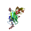
Type: D-saccharide, beta linking / Mass: 221.208 Da / Num. of mol.: 1
Source method: isolated from a genetically manipulated source
Formula: C8H15NO6
IdentifierTypeProgram
DGlcpNAcbCONDENSED IUPAC CARBOHYDRATE SYMBOLGMML 1.0
N-acetyl-b-D-glucopyranosamineCOMMON NAMEGMML 1.0
b-D-GlcpNAcIUPAC CARBOHYDRATE SYMBOLPDB-CARE 1.0
GlcNAcSNFG CARBOHYDRATE SYMBOLGMML 1.0

-
Non-polymers , 2 types, 69 molecules

#5: Chemical ChemComp-NA / SODIUM ION


Mass: 22.990 Da / Num. of mol.: 2 / Source method: obtained synthetically / Formula: Na
#6: Water ChemComp-HOH / water


Mass: 18.015 Da / Num. of mol.: 67 / Source method: isolated from a natural source / Formula: H2O

-
Details

Sequence detailsSEQUENCE DISCREPANCY, L68 IN ORIGINAL GENE BANK ENTRY IS F68

-
Experimental details

-
Experiment

ExperimentMethod: X-RAY DIFFRACTION / Number of used crystals: 1

-
Sample preparation

CrystalDensity Matthews: 3.13 Å3/Da / Density % sol: 60.34 % / Description: NONE
Crystal growTemperature: 295 K / Method: vapor diffusion, sitting drop / pH: 6.2
Details: 0.1 M TRI-SODIUM CITRATE, PH 6.2 0.1 M AMMONIUM CHLORIDE 30% (W/V) PEG 4000

-
Data collection

DiffractionMean temperature: 100 K
Diffraction sourceSource: SYNCHROTRON / Site: ESRF / Beamline: BM14 / Wavelength: 0.979
DetectorType: MARRESEARCH / Detector: CCD / Date: Nov 29, 2002
RadiationProtocol: MAD / Monochromatic (M) / Laue (L): M / Scattering type: x-ray
Radiation wavelengthWavelength: 0.979 Å / Relative weight: 1
ReflectionResolution: 2.7→30 Å / Num. obs: 10567 / % possible obs: 99.3 % / Observed criterion σ(I): 3 / Redundancy: 10.5 % / Rmerge(I) obs: 0.14 / Net I/σ(I): 12.7
Reflection shellResolution: 2.7→2.8 Å / Redundancy: 10.7 % / Rmerge(I) obs: 0.53 / Mean I/σ(I) obs: 4.1 / % possible all: 99.3

-
Processing

Software
NameVersionClassification
REFMAC5.2.0019refinement
DENZOdata reduction
SCALEPACKdata scaling
SHELXDphasing
SOLVEphasing
RESOLVEphasing
RefinementMethod to determine structure: MAD
Starting model: NONE

Resolution: 2.7→30 Å / Cor.coef. Fo:Fc: 0.934 / Cor.coef. Fo:Fc free: 0.899 / SU B: 25.656 / SU ML: 0.26 / TLS residual ADP flag: LIKELY RESIDUAL / Cross valid method: THROUGHOUT / ESU R: 0.675 / ESU R Free: 0.347 / Stereochemistry target values: MAXIMUM LIKELIHOOD / Details: HYDROGENS HAVE BEEN ADDED IN THE RIDING POSITIONS
RfactorNum. reflection% reflectionSelection details
Rfree0.275 534 4.9 %RANDOM
Rwork0.222 ---
obs0.225 10473 100 %-
Solvent computationIon probe radii: 0.8 Å / Shrinkage radii: 0.8 Å / VDW probe radii: 1.2 Å / Solvent model: MASK
Displacement parametersBiso mean: 47.97 Å2
Baniso -1Baniso -2Baniso -3
1--5.22 Å20 Å20 Å2
2--9.21 Å20 Å2
3----4 Å2
Refinement stepCycle: LAST / Resolution: 2.7→30 Å
ProteinNucleic acidLigandSolventTotal
Num. atoms2037 0 104 67 2208
Refine LS restraints
Refine-IDTypeDev idealDev ideal targetNumber
X-RAY DIFFRACTIONr_bond_refined_d0.0120.0222204
X-RAY DIFFRACTIONr_bond_other_d
X-RAY DIFFRACTIONr_angle_refined_deg1.5941.993011
X-RAY DIFFRACTIONr_angle_other_deg
X-RAY DIFFRACTIONr_dihedral_angle_1_deg7.245258
X-RAY DIFFRACTIONr_dihedral_angle_2_deg37.9223.8100
X-RAY DIFFRACTIONr_dihedral_angle_3_deg18.59815328
X-RAY DIFFRACTIONr_dihedral_angle_4_deg18.9821514
X-RAY DIFFRACTIONr_chiral_restr0.0920.2344
X-RAY DIFFRACTIONr_gen_planes_refined0.0040.021648
X-RAY DIFFRACTIONr_gen_planes_other
X-RAY DIFFRACTIONr_nbd_refined0.2380.2966
X-RAY DIFFRACTIONr_nbd_other
X-RAY DIFFRACTIONr_nbtor_refined0.3210.21477
X-RAY DIFFRACTIONr_nbtor_other
X-RAY DIFFRACTIONr_xyhbond_nbd_refined0.1660.2105
X-RAY DIFFRACTIONr_xyhbond_nbd_other
X-RAY DIFFRACTIONr_metal_ion_refined
X-RAY DIFFRACTIONr_metal_ion_other
X-RAY DIFFRACTIONr_symmetry_vdw_refined0.2340.220
X-RAY DIFFRACTIONr_symmetry_vdw_other
X-RAY DIFFRACTIONr_symmetry_hbond_refined0.1020.27
X-RAY DIFFRACTIONr_symmetry_hbond_other
X-RAY DIFFRACTIONr_symmetry_metal_ion_refined
X-RAY DIFFRACTIONr_symmetry_metal_ion_other
X-RAY DIFFRACTIONr_mcbond_it0.5041.51287
X-RAY DIFFRACTIONr_mcbond_other
X-RAY DIFFRACTIONr_mcangle_it0.96722087
X-RAY DIFFRACTIONr_mcangle_other
X-RAY DIFFRACTIONr_scbond_it1.6233952
X-RAY DIFFRACTIONr_scbond_other
X-RAY DIFFRACTIONr_scangle_it2.8584.5924
X-RAY DIFFRACTIONr_scangle_other
X-RAY DIFFRACTIONr_long_range_B_refined
X-RAY DIFFRACTIONr_long_range_B_other
X-RAY DIFFRACTIONr_rigid_bond_restr
X-RAY DIFFRACTIONr_sphericity_free
X-RAY DIFFRACTIONr_sphericity_bonded
Refinement TLS params.

Method: refined / Refine-ID: X-RAY DIFFRACTION

IDL112)L122)L132)L222)L232)L332)S11 (Å °)S12 (Å °)S13 (Å °)S21 (Å °)S22 (Å °)S23 (Å °)S31 (Å °)S32 (Å °)S33 (Å °)T112)T122)T132)T222)T232)T332)Origin x (Å)Origin y (Å)Origin z (Å)
13.00461.0449-0.91553.8229-1.64913.2679-0.0821-0.03540.1176-0.0303-0.2426-0.2276-0.29120.29670.212-0.10660.0174-0.0189-0.18960.1225-0.14739.96830.86811.134
26.36130.4746-0.34813.03880.36123.62540.0545-0.08550.031-0.15360.77180.56060.0327-0.4718-0.485-0.04860.0358-0.04180.1640.24140.09817.07532.56537.571
Refinement TLS group
IDRefine-IDRefine TLS-IDAuth asym-IDAuth seq-ID
1X-RAY DIFFRACTION1A1 - 160
2X-RAY DIFFRACTION2A161 - 259

+
About Yorodumi

-
News

-
Feb 9, 2022. New format data for meta-information of EMDB entries

New format data for meta-information of EMDB entries

  • Version 3 of the EMDB header file is now the official format.
  • The previous official version 1.9 will be removed from the archive.

Related info.:EMDB header

External links:wwPDB to switch to version 3 of the EMDB data model

-
Aug 12, 2020. Covid-19 info

Covid-19 info

URL: https://pdbjlvh1.pdbj.org/emnavi/covid19.php

New page: Covid-19 featured information page in EM Navigator.

Related info.:Covid-19 info / Mar 5, 2020. Novel coronavirus structure data

+
Mar 5, 2020. Novel coronavirus structure data

Novel coronavirus structure data

Related info.:Yorodumi Speices / Aug 12, 2020. Covid-19 info

External links:COVID-19 featured content - PDBj / Molecule of the Month (242):Coronavirus Proteases

+
Jan 31, 2019. EMDB accession codes are about to change! (news from PDBe EMDB page)

EMDB accession codes are about to change! (news from PDBe EMDB page)

  • The allocation of 4 digits for EMDB accession codes will soon come to an end. Whilst these codes will remain in use, new EMDB accession codes will include an additional digit and will expand incrementally as the available range of codes is exhausted. The current 4-digit format prefixed with “EMD-” (i.e. EMD-XXXX) will advance to a 5-digit format (i.e. EMD-XXXXX), and so on. It is currently estimated that the 4-digit codes will be depleted around Spring 2019, at which point the 5-digit format will come into force.
  • The EM Navigator/Yorodumi systems omit the EMD- prefix.

Related info.:Q: What is EMD? / ID/Accession-code notation in Yorodumi/EM Navigator

External links:EMDB Accession Codes are Changing Soon! / Contact to PDBj

+
Jul 12, 2017. Major update of PDB

Major update of PDB

  • wwPDB released updated PDB data conforming to the new PDBx/mmCIF dictionary.
  • This is a major update changing the version number from 4 to 5, and with Remediation, in which all the entries are updated.
  • In this update, many items about electron microscopy experimental information are reorganized (e.g. em_software).
  • Now, EM Navigator and Yorodumi are based on the updated data.

External links:wwPDB Remediation / Enriched Model Files Conforming to OneDep Data Standards Now Available in the PDB FTP Archive

-
Yorodumi

Thousand views of thousand structures

  • Yorodumi is a browser for structure data from EMDB, PDB, SASBDB, etc.
  • This page is also the successor to EM Navigator detail page, and also detail information page/front-end page for Omokage search.
  • The word "yorodu" (or yorozu) is an old Japanese word meaning "ten thousand". "mi" (miru) is to see.

Related info.:EMDB / PDB / SASBDB / Comparison of 3 databanks / Yorodumi Search / Aug 31, 2016. New EM Navigator & Yorodumi / Yorodumi Papers / Jmol/JSmol / Function and homology information / Changes in new EM Navigator and Yorodumi

Read more