 Movie
Movie Controller

[English] 日本語
 Yorodumi
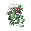
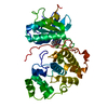
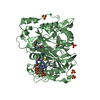
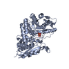
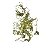
- PDB-2c6w: PENICILLIN-BINDING PROTEIN 1A (PBP-1A) FROM STREPTOCOCCUS PNEUMONIAE -
+ Open data

- Basic information
| Entry | Database: PDB / ID: 2c6w | ||||||
|---|---|---|---|---|---|---|---|
| Title | PENICILLIN-BINDING PROTEIN 1A (PBP-1A) FROM STREPTOCOCCUS PNEUMONIAE | ||||||
 Components Components | (PENICILLIN-BINDING PROTEIN 1A) x 2 | ||||||
 Keywords Keywords | PEPTIDOGLYCAN SYNTHESIS / CELL WALL / PENICILLIN-BINDING / ANTIBIOTIC RESISTANCE / CELL SHAPE / MULTIFUNCTIONAL ENZYME | ||||||
| Function / homology |  Function and homology information Function and homology informationpeptidoglycan glycosyltransferase / peptidoglycan glycosyltransferase activity / serine-type D-Ala-D-Ala carboxypeptidase / serine-type D-Ala-D-Ala carboxypeptidase activity / penicillin binding / peptidoglycan biosynthetic process / cell wall organization / regulation of cell shape / outer membrane-bounded periplasmic space / response to antibiotic ...peptidoglycan glycosyltransferase / peptidoglycan glycosyltransferase activity / serine-type D-Ala-D-Ala carboxypeptidase / serine-type D-Ala-D-Ala carboxypeptidase activity / penicillin binding / peptidoglycan biosynthetic process / cell wall organization / regulation of cell shape / outer membrane-bounded periplasmic space / response to antibiotic / proteolysis / extracellular region Similarity search - Function | ||||||
| Biological species |   STREPTOCOCCUS PNEUMONIAE (bacteria) STREPTOCOCCUS PNEUMONIAE (bacteria) | ||||||
| Method |  X-RAY DIFFRACTION / SYNCHROTRON / MOLECULAR REPLACEMENT / Resolution: 2.61 Å X-RAY DIFFRACTION / SYNCHROTRON / MOLECULAR REPLACEMENT / Resolution: 2.61 Å | ||||||
 Authors Authors | Contreras-Martel, C. / Job, V. / Di Guilmi, A.-M. / Vernet, T. / Dideberg, O. / Dessen, A. | ||||||
 Citation Citation | Journal: J.Mol.Biol. / Year: 2006 Title: Crystal Structure of Penicillin-Binding Protein 1A (Pbp1A) Reveals a Mutational Hotspot Implicated in Beta-Lactam Resistance in Streptococcus Pneumoniae. Authors: Contreras-Martel, C. / Job, V. / Di Guilmi, A.-M. / Vernet, T. / Dideberg, O. / Dessen, A. #1: Journal: Acta Crystallogr.,Sect.D / Year: 2003 Title: Structural Studies of the Transpeptidase Domain of Pbp1A from Streptococcus Pneumoniae Authors: Job, V. / Di Guilmi, A.-M. / Martin, L. / Vernet, T. / Dideberg, O. / Dessen, A. #2: Journal: J.Bacteriol. / Year: 1998 Title: Identification, Purification, and Charactherization of Transpeptidase and Glycosyltransferase Domains of Streptococcus Pneumoniae Penicillin-Binding Protein 1A Authors: Di Guilmi, A.-M. / Mouz, N. / Andrieu, J.-P. / Hoskins, J. / Jaskunas, S.R. / Gagnon, J. / Dideberg, O. / Vernet, T. | ||||||
| History |
|
- Structure visualization
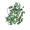
| Structure viewer | Molecule: MolmilJmol/JSmol |
|---|
- Downloads & links
-Download
| PDBx/mmCIF format | 2c6w.cif.gz | 94 KB | Display | PDBx/mmCIF format |
|---|---|---|---|---|
| PDB format | pdb2c6w.ent.gz | 70.4 KB | Display | PDB format |
| PDBx/mmJSON format | 2c6w.json.gz | Tree view | PDBx/mmJSON format | |
| Others |  Other downloads Other downloads |
-Validation report
| Summary document | 2c6w_validation.pdf.gz | 435.7 KB | Display | wwPDB validaton report |
|---|---|---|---|---|
| Full document | 2c6w_full_validation.pdf.gz | 449.5 KB | Display | |
| Data in XML | 2c6w_validation.xml.gz | 18.5 KB | Display | |
| Data in CIF | 2c6w_validation.cif.gz | 24.4 KB | Display | |
| Arichive directory | https://data.pdbj.org/pub/pdb/validation_reports/c6/2c6wftp://data.pdbj.org/pub/pdb/validation_reports/c6/2c6w | HTTPS FTP |
-Related structure data
| Related structure data | 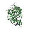 2c5wSC S: Starting model for refinement C: citing same article ( |
|---|---|
| Similar structure data |


-Links
 PDBj
PDBj
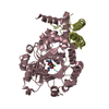
- Assembly
| Deposited unit | 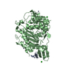
| ||||||||
|---|---|---|---|---|---|---|---|---|---|
| 1 |
| ||||||||
| Unit cell |
| ||||||||
| Details | PBP-1A IS A MONOMER IN SOLUTION, BUT SINCE IN THIS ENTRYTHERE IS A CLEAVED PEPTIDE FROM THE SAME PROTEIN (CHAIN A)ASSOCIATED WITH THE TRANSPEPTIDASE DOMAIN OFPBP-1A (CHAIN B), THE ENTRY IS MARKED AS DIMERIC. |
-Components
| #1: Protein/peptide | Mass: 1825.027 Da / Num. of mol.: 1 / Fragment: GLYCOSYLTRANSFERASE DOMAIN, RESIDUES 51-66 Source method: isolated from a genetically manipulated source Source: (gene. exp.) STREPTOCOCCUS PNEUMONIAE (bacteria) / Strain: R6 / Plasmid: PJAH143 / Production host: | ||||||||
|---|---|---|---|---|---|---|---|---|---|
| #2: Protein | Mass: 43078.492 Da / Num. of mol.: 1 / Fragment: TRANSPEPTIDASE DOMAIN, RESIDUES 267-650 Source method: isolated from a genetically manipulated source Source: (gene. exp.) STREPTOCOCCUS PNEUMONIAE (bacteria) / Strain: R6 / Plasmid: PJAH143 / Production host: | ||||||||
| #3: Chemical |   Mass: 65.409 Da / Num. of mol.: 3 / Source method: obtained synthetically / Formula: Zn Mass: 65.409 Da / Num. of mol.: 3 / Source method: obtained synthetically / Formula: Zn#4: Chemical | ChemComp-CL /   Mass: 35.453 Da / Num. of mol.: 4 / Source method: obtained synthetically / Formula: Cl Mass: 35.453 Da / Num. of mol.: 4 / Source method: obtained synthetically / Formula: Cl#5: Water | ChemComp-HOH / |  Mass: 18.015 Da / Num. of mol.: 35 / Source method: isolated from a natural source / Formula: H2O Mass: 18.015 Da / Num. of mol.: 35 / Source method: isolated from a natural source / Formula: H2OCompound details | CELL WALL FORMATION | Sequence details | EXTRA PEPTIDE FROM THE GLYLOSYLTRANSFERASE DOMAIN, RESIDUES 51-66. THE SEQUENCE IN THE SEQRES ...EXTRA PEPTIDE FROM THE GLYLOSYLTR | |
-Experimental details
-Experiment
| Experiment | Method: X-RAY DIFFRACTION / Number of used crystals: 1 |
|---|
- Sample preparation
| Crystal | Density Matthews: 3.03 Å3/Da / Density % sol: 57.79 % |
|---|---|
| Crystal grow | pH: 7 Details: 13% PEG1000, 50MM NACL, 5MM ZNSO4, 50MM TRIS PH 7.0 |
-Data collection
| Diffraction | Mean temperature: 100 K |
|---|---|
| Diffraction source | Source: SYNCHROTRON / Site: ESRF  / Beamline: ID13 / Wavelength: 0.964 / Beamline: ID13 / Wavelength: 0.964 |
| Detector | Type: MARRESEARCH / Detector: CCD / Date: Mar 24, 2001 |
| Radiation | Protocol: SINGLE WAVELENGTH / Monochromatic (M) / Laue (L): M / Scattering type: x-ray |
| Radiation wavelength | Wavelength: 0.964 Å / Relative weight: 1 |
| Reflection | Resolution: 2.61→50 Å / Num. obs: 15084 / % possible obs: 91.9 % / Observed criterion σ(I): 2 / Redundancy: 3.4 % / Biso Wilson estimate: 72.7 Å2 / Rsym value: 0.08 / Net I/σ(I): 10.2 |
| Reflection shell | Resolution: 2.61→2.77 Å / Redundancy: 3 % / Mean I/σ(I) obs: 3.5 / Rsym value: 0.3 / % possible all: 72.4 |
- Processing
| Software |
| ||||||||||||||||||||||||||||||||||||||||||||||||||||||||||||||||||||||||||||||||
|---|---|---|---|---|---|---|---|---|---|---|---|---|---|---|---|---|---|---|---|---|---|---|---|---|---|---|---|---|---|---|---|---|---|---|---|---|---|---|---|---|---|---|---|---|---|---|---|---|---|---|---|---|---|---|---|---|---|---|---|---|---|---|---|---|---|---|---|---|---|---|---|---|---|---|---|---|---|---|---|---|---|
| Refinement | Method to determine structure: MOLECULAR REPLACEMENT Starting model: PDB ENTRY 2C5W Resolution: 2.61→44.99 Å / Rfactor Rfree error: 0.01 / Data cutoff high absF: 3749779.78 / Isotropic thermal model: RESTRAINED / Cross valid method: THROUGHOUT / σ(F): 0 / Stereochemistry target values: MAXIMUM LIKELIHOOD
| ||||||||||||||||||||||||||||||||||||||||||||||||||||||||||||||||||||||||||||||||
| Solvent computation | Solvent model: FLAT MODEL / Bsol: 68.5283 Å2 / ksol: 0.349369 e/Å3 | ||||||||||||||||||||||||||||||||||||||||||||||||||||||||||||||||||||||||||||||||
| Displacement parameters | Biso mean: 68.5 Å2
| ||||||||||||||||||||||||||||||||||||||||||||||||||||||||||||||||||||||||||||||||
| Refine analyze |
| ||||||||||||||||||||||||||||||||||||||||||||||||||||||||||||||||||||||||||||||||
| Refinement step | Cycle: LAST / Resolution: 2.61→44.99 Å
| ||||||||||||||||||||||||||||||||||||||||||||||||||||||||||||||||||||||||||||||||
| Refine LS restraints |
| ||||||||||||||||||||||||||||||||||||||||||||||||||||||||||||||||||||||||||||||||
| LS refinement shell | Resolution: 2.61→2.77 Å / Rfactor Rfree error: 0.045 / Total num. of bins used: 6
| ||||||||||||||||||||||||||||||||||||||||||||||||||||||||||||||||||||||||||||||||
| Xplor file |
|
