[English] 日本語
Yorodumi
- PDB-2c4r: Catalytic domain of E. coli RNase E -

+
Open data


ID or keywords:

Loading...

-
Basic information

Entry
Database: PDB / ID: 2c4r
TitleCatalytic domain of E. coli RNase E
Components
  • RIBONUCLEASE E
  • SSRNA MOLECULE: 5'-R(*AP*CP*AP*GP*UP*AP*UP*UP*UP*GP)-3'
KeywordsHYDROLASE / RNA BINDING / RNA TURNOVER / RNA PROCESSING / ENDONUCLEASE / NUCLEASE
Function / homology
Function and homology information


regulation of RNA helicase activity / rRNA 5'-end processing / ribonuclease E / ribonuclease E activity / bacterial degradosome / endoribonuclease complex / DEAD/H-box RNA helicase binding / 7S RNA binding / RNA catabolic process / tRNA processing ...regulation of RNA helicase activity / rRNA 5'-end processing / ribonuclease E / ribonuclease E activity / bacterial degradosome / endoribonuclease complex / DEAD/H-box RNA helicase binding / 7S RNA binding / RNA catabolic process / tRNA processing / mRNA catabolic process / RNA nuclease activity / RNA processing / RNA endonuclease activity / cytoplasmic side of plasma membrane / rRNA processing / protein complex oligomerization / protein homotetramerization / tRNA binding / molecular adaptor activity / rRNA binding / magnesium ion binding / RNA binding / zinc ion binding / identical protein binding / membrane / cytoplasm
Similarity search - Function
Ribonuclease E, catalytic domain / Hypothetical Protein Ychn; Chain: A, / Polyribonucleotide phosphorylase C-terminal / Polyribonucleotide phosphorylase C terminal / : / RNase E/G, Thioredoxin-like domain / Ribonuclease E/G / RNA-binding protein AU-1/Ribonuclease E/G / Ribonuclease E / Ribonuclease E/G family ...Ribonuclease E, catalytic domain / Hypothetical Protein Ychn; Chain: A, / Polyribonucleotide phosphorylase C-terminal / Polyribonucleotide phosphorylase C terminal / : / RNase E/G, Thioredoxin-like domain / Ribonuclease E/G / RNA-binding protein AU-1/Ribonuclease E/G / Ribonuclease E / Ribonuclease E/G family / S1 domain profile. / Ribosomal protein S1-like RNA-binding domain / S1 RNA binding domain / S1 domain / Nucleic acid-binding, OB-fold / 3-Layer(aba) Sandwich / Alpha Beta
Similarity search - Domain/homology
RNA / Ribonuclease E
Similarity search - Component
Biological speciesESCHERICHIA COLI (E. coli)
MethodX-RAY DIFFRACTION / SYNCHROTRON / MOLECULAR REPLACEMENT / Resolution: 3.6 Å
AuthorsMarcaida, M.J. / Callaghan, A.J. / Luisi, B.F.
CitationJournal: Nature / Year: 2005
Title: Structure of E. Coli Rnase E Catalytic Domain and Implications for RNA Processing and Turnover
Authors: Callaghan, A.J. / Marcaida, M.J. / Stead, J.A. / McDowall, K.J. / Scott, W.G. / Luisi, B.F.
History
DepositionOct 21, 2005Deposition site: PDBE / Processing site: PDBE
Revision 1.0Oct 25, 2005Provider: repository / Type: Initial release
Revision 1.1Jul 13, 2011Group: Advisory / Refinement description / Version format compliance
Revision 1.2Apr 4, 2018Group: Data collection / Database references / Category: citation_author / Item: _citation_author.name
Revision 1.3Dec 13, 2023Group: Data collection / Database references ...Data collection / Database references / Derived calculations / Other / Refinement description
Category: chem_comp_atom / chem_comp_bond ...chem_comp_atom / chem_comp_bond / database_2 / pdbx_database_status / pdbx_initial_refinement_model / pdbx_struct_conn_angle / struct_conn
Item: _database_2.pdbx_DOI / _database_2.pdbx_database_accession ..._database_2.pdbx_DOI / _database_2.pdbx_database_accession / _pdbx_database_status.status_code_sf / _pdbx_struct_conn_angle.ptnr1_auth_asym_id / _pdbx_struct_conn_angle.ptnr1_auth_comp_id / _pdbx_struct_conn_angle.ptnr1_auth_seq_id / _pdbx_struct_conn_angle.ptnr1_label_asym_id / _pdbx_struct_conn_angle.ptnr1_label_atom_id / _pdbx_struct_conn_angle.ptnr1_label_comp_id / _pdbx_struct_conn_angle.ptnr1_label_seq_id / _pdbx_struct_conn_angle.ptnr1_symmetry / _pdbx_struct_conn_angle.ptnr3_auth_asym_id / _pdbx_struct_conn_angle.ptnr3_auth_comp_id / _pdbx_struct_conn_angle.ptnr3_auth_seq_id / _pdbx_struct_conn_angle.ptnr3_label_asym_id / _pdbx_struct_conn_angle.ptnr3_label_atom_id / _pdbx_struct_conn_angle.ptnr3_label_comp_id / _pdbx_struct_conn_angle.ptnr3_label_seq_id / _pdbx_struct_conn_angle.ptnr3_symmetry / _pdbx_struct_conn_angle.value / _struct_conn.pdbx_dist_value / _struct_conn.ptnr1_auth_comp_id / _struct_conn.ptnr1_auth_seq_id / _struct_conn.ptnr1_label_asym_id / _struct_conn.ptnr1_label_atom_id / _struct_conn.ptnr1_label_comp_id / _struct_conn.ptnr1_label_seq_id / _struct_conn.ptnr1_symmetry / _struct_conn.ptnr2_auth_asym_id / _struct_conn.ptnr2_auth_comp_id / _struct_conn.ptnr2_auth_seq_id / _struct_conn.ptnr2_label_asym_id / _struct_conn.ptnr2_label_atom_id / _struct_conn.ptnr2_label_comp_id / _struct_conn.ptnr2_label_seq_id / _struct_conn.ptnr2_symmetry
Remark 700 
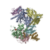
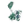
SHEET THE SHEET STRUCTURE OF THIS MOLECULE IS BIFURCATED. IN ORDER TO REPRESENT THIS FEATURE IN ... SHEET THE SHEET STRUCTURE OF THIS MOLECULE IS BIFURCATED. IN ORDER TO REPRESENT THIS FEATURE IN THE SHEET RECORDS BELOW, TWO SHEETS ARE DEFINED.

-
Structure visualization

Structure viewerMolecule:
MolmilJmol/JSmol

Downloads & links

-
Assembly

Deposited unit
L: RIBONUCLEASE E
R: SSRNA MOLECULE: 5'-R(*AP*CP*AP*GP*UP*AP*UP*UP*UP*GP)-3'
hetero molecules


Theoretical massNumber of molelcules
Total (without water)61,4925
Polymers61,3782
Non-polymers1143
Water1086
1
L: RIBONUCLEASE E
R: SSRNA MOLECULE: 5'-R(*AP*CP*AP*GP*UP*AP*UP*UP*UP*GP)-3'
hetero molecules

L: RIBONUCLEASE E
R: SSRNA MOLECULE: 5'-R(*AP*CP*AP*GP*UP*AP*UP*UP*UP*GP)-3'
hetero molecules

L: RIBONUCLEASE E
R: SSRNA MOLECULE: 5'-R(*AP*CP*AP*GP*UP*AP*UP*UP*UP*GP)-3'
hetero molecules

L: RIBONUCLEASE E
R: SSRNA MOLECULE: 5'-R(*AP*CP*AP*GP*UP*AP*UP*UP*UP*GP)-3'
hetero molecules


Theoretical massNumber of molelcules
Total (without water)245,96820
Polymers245,5128
Non-polymers45612
Water1448
TypeNameSymmetry operationNumber
identity operation1_555x,y,z1
crystal symmetry operation4_545-x,-y-1,z1
crystal symmetry operation12_545x,x-y-1,-z+1/31
crystal symmetry operation9_555-x,-x+y,-z+1/31
MethodPQS
Unit cell
Length a, b, c (Å)196.586, 196.586, 140.766
Angle α, β, γ (deg.)90.00, 90.00, 120.00
Int Tables number180
Space group name H-MP6222
Components on special symmetry positions
IDModelComponents
11L-1512-

ZN

-
Components

#1: Protein RIBONUCLEASE E / RNASE E


Mass: 58215.133 Da / Num. of mol.: 1 / Fragment: CATALYTIC DOMAIN, RESIDUES 1-510
Source method: isolated from a genetically manipulated source
Source: (gene. exp.) ESCHERICHIA COLI (E. coli) / Production host: ESCHERICHIA COLI (E. coli) / Strain (production host): BL21(DE3)
References: UniProt: P21513, Hydrolases; Acting on ester bonds; Phosphoric-diester hydrolases
#2: RNA chain SSRNA MOLECULE: 5'-R(*AP*CP*AP*GP*UP*AP*UP*UP*UP*GP)-3'


Mass: 3162.916 Da / Num. of mol.: 1 / Source method: obtained synthetically / Source: (synth.) ESCHERICHIA COLI (E. coli)
#3: Chemical ChemComp-MG / MAGNESIUM ION


Mass: 24.305 Da / Num. of mol.: 2 / Source method: obtained synthetically / Formula: Mg
#4: Chemical ChemComp-ZN / ZINC ION


Mass: 65.409 Da / Num. of mol.: 1 / Source method: obtained synthetically / Formula: Zn
#5: Water ChemComp-HOH / water


Mass: 18.015 Da / Num. of mol.: 6 / Source method: isolated from a natural source / Formula: H2O
Compound detailsHAS A ROLE IN THE MATURATION OF 5S RRNA FROM ITS PRECURSORS FROM ALL THE RRNA GENES

-
Experimental details

-
Experiment

ExperimentMethod: X-RAY DIFFRACTION / Number of used crystals: 1

-
Sample preparation

CrystalDensity Matthews: 7.1 Å3/Da / Density % sol: 82 %
Crystal growpH: 8
Details: CRYSTALLIZATION CONDITIONS: CRYSTALS OF THE RNASE E CATALYTIC DOMAIN/ RNA COMPLEX APPEARED AFTER TWO TO FOUR WEEKS IN 5 TO 20 % WT/V POLYETHYLENE GLYCOL 8,000, 0.1 M TRIS PH 7.5 TO 8.0, AND ...Details: CRYSTALLIZATION CONDITIONS: CRYSTALS OF THE RNASE E CATALYTIC DOMAIN/ RNA COMPLEX APPEARED AFTER TWO TO FOUR WEEKS IN 5 TO 20 % WT/V POLYETHYLENE GLYCOL 8,000, 0.1 M TRIS PH 7.5 TO 8.0, AND 10 TO 50 MM MAGNESIUM FORMATE AT 20OC

-
Data collection

DiffractionMean temperature: 100 K
Diffraction sourceSource: SYNCHROTRON / Site: ALS / Type: ALS / Wavelength: 1.1
DetectorType: ADSC CCD / Detector: CCD
RadiationProtocol: SINGLE WAVELENGTH / Monochromatic (M) / Laue (L): M / Scattering type: x-ray
Radiation wavelengthWavelength: 1.1 Å / Relative weight: 1
ReflectionResolution: 3.6→25 Å / Num. obs: 19065 / % possible obs: 99.8 % / Observed criterion σ(I): 0 / Redundancy: 16 % / Rmerge(I) obs: 0.13 / Net I/σ(I): 23
Reflection shellResolution: 3.6→3.73 Å / Redundancy: 10 % / Rmerge(I) obs: 0.64 / Mean I/σ(I) obs: 209 / % possible all: 97.7

-
Processing

Software
NameVersionClassification
REFMAC5.2.0005refinement
HKL-2000data reduction
SCALEPACKdata scaling
RefinementMethod to determine structure: MOLECULAR REPLACEMENT
Starting model: PDB ENTRY 2BX2
Resolution: 3.6→25 Å / Cor.coef. Fo:Fc: 0.862 / Cor.coef. Fo:Fc free: 0.838 / SU B: 42.384 / SU ML: 0.306 / TLS residual ADP flag: LIKELY RESIDUAL / Cross valid method: THROUGHOUT / ESU R: 1.355 / ESU R Free: 0.584 / Stereochemistry target values: MAXIMUM LIKELIHOOD
Details: HYDROGENS HAVE BEEN ADDED IN THE RIDING POSITIONS. DISORDERED REGIONS WERE REMOVED FROM THE MODEL
RfactorNum. reflection% reflectionSelection details
Rfree0.347 973 5.1 %RANDOM
Rwork0.319 ---
obs0.32 17938 99.7 %-
Solvent computationIon probe radii: 0.8 Å / Shrinkage radii: 0.8 Å / VDW probe radii: 1.2 Å / Solvent model: BABINET MODEL WITH MASK
Displacement parametersBiso mean: 59.1 Å2
Baniso -1Baniso -2Baniso -3
1-4.22 Å22.11 Å20 Å2
2--4.22 Å20 Å2
3----6.33 Å2
Refinement stepCycle: LAST / Resolution: 3.6→25 Å
ProteinNucleic acidLigandSolventTotal
Num. atoms3557 212 3 6 3778
Refine LS restraints
Refine-IDTypeDev idealDev ideal targetNumber
X-RAY DIFFRACTIONr_bond_refined_d0.0160.0213855
X-RAY DIFFRACTIONr_bond_other_d
X-RAY DIFFRACTIONr_angle_refined_deg1.9752.0375300
X-RAY DIFFRACTIONr_angle_other_deg
X-RAY DIFFRACTIONr_dihedral_angle_1_deg14.2455487
X-RAY DIFFRACTIONr_dihedral_angle_2_deg40.72623.172145
X-RAY DIFFRACTIONr_dihedral_angle_3_deg28.26615534
X-RAY DIFFRACTIONr_dihedral_angle_4_deg24.5221532
X-RAY DIFFRACTIONr_chiral_restr0.1180.2637
X-RAY DIFFRACTIONr_gen_planes_refined0.0120.022854
X-RAY DIFFRACTIONr_gen_planes_other
X-RAY DIFFRACTIONr_nbd_refined0.4060.32298
X-RAY DIFFRACTIONr_nbd_other
X-RAY DIFFRACTIONr_nbtor_refined0.3770.52655
X-RAY DIFFRACTIONr_nbtor_other
X-RAY DIFFRACTIONr_xyhbond_nbd_refined0.3590.5295
X-RAY DIFFRACTIONr_xyhbond_nbd_other
X-RAY DIFFRACTIONr_metal_ion_refined
X-RAY DIFFRACTIONr_metal_ion_other
X-RAY DIFFRACTIONr_symmetry_vdw_refined0.6570.382
X-RAY DIFFRACTIONr_symmetry_vdw_other
X-RAY DIFFRACTIONr_symmetry_hbond_refined0.3540.513
X-RAY DIFFRACTIONr_symmetry_hbond_other
X-RAY DIFFRACTIONr_symmetry_metal_ion_refined
X-RAY DIFFRACTIONr_symmetry_metal_ion_other
X-RAY DIFFRACTIONr_mcbond_it3.21122460
X-RAY DIFFRACTIONr_mcbond_other
X-RAY DIFFRACTIONr_mcangle_it5.49433864
X-RAY DIFFRACTIONr_mcangle_other
X-RAY DIFFRACTIONr_scbond_it3.37321554
X-RAY DIFFRACTIONr_scbond_other
X-RAY DIFFRACTIONr_scangle_it5.40131433
X-RAY DIFFRACTIONr_scangle_other
X-RAY DIFFRACTIONr_long_range_B_refined
X-RAY DIFFRACTIONr_long_range_B_other
X-RAY DIFFRACTIONr_rigid_bond_restr
X-RAY DIFFRACTIONr_sphericity_free
X-RAY DIFFRACTIONr_sphericity_bonded
LS refinement shellResolution: 3.6→3.7 Å / Total num. of bins used: 20 /
RfactorNum. reflection
Rfree0.36 67
Rwork0.31 1231
Refinement TLS params.Method: refined / Origin x: 72.5883 Å / Origin y: -62.5588 Å / Origin z: 10.2861 Å
111213212223313233
T-0.32 Å2-0.17 Å20.0078 Å2--0.151 Å20.051 Å2---0.063 Å2
L1.8207 °20.3982 °2-1.22 °2-0.4304 °2-0.1712 °2--3.8987 °2
S0.1962 Å °0.4367 Å °0.6301 Å °0.0112 Å °0.0613 Å °0.1876 Å °-0.5989 Å °-0.0531 Å °-0.2576 Å °
Refinement TLS group
IDRefine-IDRefine TLS-IDAuth asym-IDAuth seq-ID
1X-RAY DIFFRACTION1L-6 - 503
2X-RAY DIFFRACTION1R1 - 10

+
About Yorodumi

-
News

-
Feb 9, 2022. New format data for meta-information of EMDB entries

New format data for meta-information of EMDB entries

  • Version 3 of the EMDB header file is now the official format.
  • The previous official version 1.9 will be removed from the archive.

Related info.:EMDB header

External links:wwPDB to switch to version 3 of the EMDB data model

-
Aug 12, 2020. Covid-19 info

Covid-19 info

URL: https://pdbjlvh1.pdbj.org/emnavi/covid19.php

New page: Covid-19 featured information page in EM Navigator.

Related info.:Covid-19 info / Mar 5, 2020. Novel coronavirus structure data

+
Mar 5, 2020. Novel coronavirus structure data

Novel coronavirus structure data

Related info.:Yorodumi Speices / Aug 12, 2020. Covid-19 info

External links:COVID-19 featured content - PDBj / Molecule of the Month (242):Coronavirus Proteases

+
Jan 31, 2019. EMDB accession codes are about to change! (news from PDBe EMDB page)

EMDB accession codes are about to change! (news from PDBe EMDB page)

  • The allocation of 4 digits for EMDB accession codes will soon come to an end. Whilst these codes will remain in use, new EMDB accession codes will include an additional digit and will expand incrementally as the available range of codes is exhausted. The current 4-digit format prefixed with “EMD-” (i.e. EMD-XXXX) will advance to a 5-digit format (i.e. EMD-XXXXX), and so on. It is currently estimated that the 4-digit codes will be depleted around Spring 2019, at which point the 5-digit format will come into force.
  • The EM Navigator/Yorodumi systems omit the EMD- prefix.

Related info.:Q: What is EMD? / ID/Accession-code notation in Yorodumi/EM Navigator

External links:EMDB Accession Codes are Changing Soon! / Contact to PDBj

+
Jul 12, 2017. Major update of PDB

Major update of PDB

  • wwPDB released updated PDB data conforming to the new PDBx/mmCIF dictionary.
  • This is a major update changing the version number from 4 to 5, and with Remediation, in which all the entries are updated.
  • In this update, many items about electron microscopy experimental information are reorganized (e.g. em_software).
  • Now, EM Navigator and Yorodumi are based on the updated data.

External links:wwPDB Remediation / Enriched Model Files Conforming to OneDep Data Standards Now Available in the PDB FTP Archive

-
Yorodumi

Thousand views of thousand structures

  • Yorodumi is a browser for structure data from EMDB, PDB, SASBDB, etc.
  • This page is also the successor to EM Navigator detail page, and also detail information page/front-end page for Omokage search.
  • The word "yorodu" (or yorozu) is an old Japanese word meaning "ten thousand". "mi" (miru) is to see.

Related info.:EMDB / PDB / SASBDB / Comparison of 3 databanks / Yorodumi Search / Aug 31, 2016. New EM Navigator & Yorodumi / Yorodumi Papers / Jmol/JSmol / Function and homology information / Changes in new EM Navigator and Yorodumi

Read more