Mass: 18.015 Da / Num. of mol.: 55 / Source method: isolated from a natural source / Formula: H2O
Sequence details
AUTHORS SEQUENCE NOT YET DEPOSITED IN SEQUENCE DATABASE. RESIDUE THR 3 HAS BEEN MUTATED TO VAL 3. ...AUTHORS SEQUENCE NOT YET DEPOSITED IN SEQUENCE DATABASE. RESIDUE THR 3 HAS BEEN MUTATED TO VAL 3. THE GENBANK REFERENCE CODE FOR THIS ENTRY IS GQ232759.
-
Experimental details
-
Experiment
Experiment
Method: X-RAY DIFFRACTION / Number of used crystals: 1
-
Sample preparation
Crystal
Density Matthews: 3.3 Å3/Da / Density % sol: 62.6 %
Resolution: 2.6→92.45 Å / Cor.coef. Fo:Fc: 0.944 / Cor.coef. Fo:Fc free: 0.889 / SU B: 11.161 / SU ML: 0.23 / Cross valid method: THROUGHOUT / ESU R: 0.47 / ESU R Free: 0.311 / Stereochemistry target values: MAXIMUM LIKELIHOOD / Details: HYDROGENS HAVE BEEN ADDED IN THE RIDING POSITIONS.
Rfactor
Num. reflection
% reflection
Selection details
Rfree
0.271
912
5.1 %
RANDOM
Rwork
0.202
-
-
-
obs
0.205
16965
99.9 %
-
Solvent computation
Ion probe radii: 0.8 Å / Shrinkage radii: 0.8 Å / VDW probe radii: 1.2 Å / Solvent model: MASK
 Movie
Movie Controller

 Yorodumi
 Open data
 Basic information
 Components
 Keywords
 Function and homology information
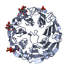
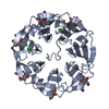
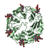
 PSATHYRELLA VELUTINA (fungus)
 X-RAY DIFFRACTION /
  Authors
 Citation
 Structure visualization
 Downloads & links
 Other downloads


 PDBj
PDBj
 Assembly

 Mass: 40.078 Da / Num. of mol.: 1 / Source method: obtained synthetically / Formula: Ca
 Type: D-saccharide, beta linking / Mass: 221.208 Da / Num. of mol.: 6
 Mass: 96.063 Da / Num. of mol.: 4 / Source method: obtained synthetically / Formula: SO4
 Mass: 18.015 Da / Num. of mol.: 55 / Source method: isolated from a natural source / Formula: H2O
 Sample preparation
 / Beamline: ID23-1 / Wavelength: 0.953
  Processing