 Movie
Movie Controller

[English] 日本語
 Yorodumi
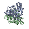
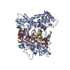
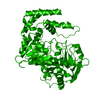
- PDB-2c2g: Crystal structure of Threonine Synthase from Arabidopsis thaliana... -
+ Open data

- Basic information
| Entry | Database: PDB / ID: 2c2g | ||||||
|---|---|---|---|---|---|---|---|
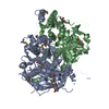
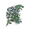
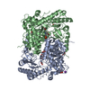
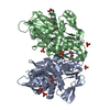
| Title | Crystal structure of Threonine Synthase from Arabidopsis thaliana in complex with its cofactor pyridoxal phosphate | ||||||
 Components Components | THREONINE SYNTHASE | ||||||
 Keywords Keywords | LYASE / SYNTHASE / THREONINE BIOSYNTHESIS / PYRIDOXAL PHOSPHATE / AMINO-ACID BIOSYNTHESIS | ||||||
| Function / homology |  Function and homology information Function and homology informationmeristem maintenance / threonine synthase / threonine synthase activity / L-threonine biosynthetic process / L-cysteine biosynthetic process / chloroplast stroma / chloroplast / pyridoxal phosphate binding / plasma membrane / cytoplasm Similarity search - Function | ||||||
| Biological species |  | ||||||
| Method |  X-RAY DIFFRACTION / MOLECULAR REPLACEMENT / Resolution: 2.61 Å X-RAY DIFFRACTION / MOLECULAR REPLACEMENT / Resolution: 2.61 Å | ||||||
 Authors Authors | Mas-Droux, C. / Biou, V. / Dumas, R. | ||||||
 Citation Citation | Journal: J.Biol.Chem. / Year: 2006 Title: Allosteric Threonine Synthase: Reorganization of the Pyridoxal Phosphate Site Upon Asymmetric Activation Through S-Adenosylmethionine Binding to a Novel Site. Authors: Mas-Droux, C. / Biou, V. / Dumas, R. | ||||||
| History |
|
- Structure visualization
| Structure viewer | Molecule: MolmilJmol/JSmol |
|---|
- Downloads & links
-Download
| PDBx/mmCIF format | 2c2g.cif.gz | 183 KB | Display | PDBx/mmCIF format |
|---|---|---|---|---|
| PDB format | pdb2c2g.ent.gz | 144.9 KB | Display | PDB format |
| PDBx/mmJSON format | 2c2g.json.gz | Tree view | PDBx/mmJSON format | |
| Others |  Other downloads Other downloads |
-Validation report
| Arichive directory | https://data.pdbj.org/pub/pdb/validation_reports/c2/2c2gftp://data.pdbj.org/pub/pdb/validation_reports/c2/2c2g | HTTPS FTP |
|---|
-Related structure data
| Related structure data |  2c2bC  1e5xS S: Starting model for refinement C: citing same article ( |
|---|---|
| Similar structure data |

-Links
 PDBj
PDBj
- Assembly
| Deposited unit | 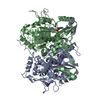
| ||||||||||||||||||
|---|---|---|---|---|---|---|---|---|---|---|---|---|---|---|---|---|---|---|---|
| 1 |
| ||||||||||||||||||
| Unit cell |
| ||||||||||||||||||
| Noncrystallographic symmetry (NCS) | NCS domain:
NCS domain segments: Component-ID: 1 / Ens-ID: 1 / Beg auth comp-ID: VAL / Beg label comp-ID: VAL / End auth comp-ID: LEU / End label comp-ID: LEU / Refine code: 4 / Auth seq-ID: 36 - 479 / Label seq-ID: 36 - 479
NCS oper: (Code: given Matrix: (0.93402, -0.16235, 0.31818), Vector: |
-Components
| #1: Protein | Mass: 53385.590 Da / Num. of mol.: 2 Source method: isolated from a genetically manipulated source Details: RESIDUE 163 IS A LYSINE BOUND TO PYRIDOXAL PHOSPHATE VIA A SCHIFF BASE Source: (gene. exp.)  #2: Chemical |   Mass: 247.142 Da / Num. of mol.: 2 / Source method: obtained synthetically / Formula: C8H10NO6P Mass: 247.142 Da / Num. of mol.: 2 / Source method: obtained synthetically / Formula: C8H10NO6P#3: Water | ChemComp-HOH / |  Mass: 18.015 Da / Num. of mol.: 121 / Source method: isolated from a natural source / Formula: H2O Mass: 18.015 Da / Num. of mol.: 121 / Source method: isolated from a natural source / Formula: H2OCompound details | HYDROLYSES O-PHOSPHO-L-HOMOSERINE INTO L-THREONINE AND PHOSPHATE. OTHER_DETAILS: RESIDUE 163 IS A ...HYDROLYSES | |
|---|
-Experimental details
-Experiment
| Experiment | Method: X-RAY DIFFRACTION / Number of used crystals: 1 |
|---|
- Sample preparation
| Crystal | Density Matthews: 2.33 Å3/Da / Density % sol: 46.85 % |
|---|---|
| Crystal grow | pH: 6.5 Details: 1 M LICL, 13% (W/V) PEG 6000, 5 MM DTT, 0.1 M MES-KOH, pH 6.50 |
-Data collection
| Diffraction | Mean temperature: 100 K |
|---|---|
| Diffraction source | Source: ROTATING ANODE / Type: RIGAKU RU200 / Wavelength: 1.5418 |
| Detector | Type: MAR scanner 345 mm plate / Detector: IMAGE PLATE / Date: Feb 20, 2000 / Details: MIRRORS |
| Radiation | Protocol: SINGLE WAVELENGTH / Monochromatic (M) / Laue (L): M / Scattering type: x-ray |
| Radiation wavelength | Wavelength: 1.5418 Å / Relative weight: 1 |
| Reflection | Resolution: 2.6→20 Å / Num. obs: 25774 / % possible obs: 95.5 % / Observed criterion σ(I): 0 / Redundancy: 2 % / Rmerge(I) obs: 0.05 / Net I/σ(I): 6.8 |
| Reflection shell | Resolution: 2.61→2.75 Å / Redundancy: 2 % / Rmerge(I) obs: 0.29 / Mean I/σ(I) obs: 2.5 / % possible all: 93.5 |
- Processing
| Software |
| ||||||||||||||||||||||||||||||||||||||||||||||||||||||||||||||||||||||||||||||||||||||||||||||||||||||||||||||||||||||||||||||||||||||||||||||||||||||||||||||||||||||||||||||||||||||
|---|---|---|---|---|---|---|---|---|---|---|---|---|---|---|---|---|---|---|---|---|---|---|---|---|---|---|---|---|---|---|---|---|---|---|---|---|---|---|---|---|---|---|---|---|---|---|---|---|---|---|---|---|---|---|---|---|---|---|---|---|---|---|---|---|---|---|---|---|---|---|---|---|---|---|---|---|---|---|---|---|---|---|---|---|---|---|---|---|---|---|---|---|---|---|---|---|---|---|---|---|---|---|---|---|---|---|---|---|---|---|---|---|---|---|---|---|---|---|---|---|---|---|---|---|---|---|---|---|---|---|---|---|---|---|---|---|---|---|---|---|---|---|---|---|---|---|---|---|---|---|---|---|---|---|---|---|---|---|---|---|---|---|---|---|---|---|---|---|---|---|---|---|---|---|---|---|---|---|---|---|---|---|---|
| Refinement | Method to determine structure: MOLECULAR REPLACEMENT Starting model: PDB ENTRY 1E5X Resolution: 2.61→20.63 Å / Cor.coef. Fo:Fc: 0.947 / Cor.coef. Fo:Fc free: 0.898 / SU B: 27.272 / SU ML: 0.28 / TLS residual ADP flag: UNVERIFIED / Cross valid method: THROUGHOUT / ESU R Free: 0.376 / Stereochemistry target values: MAXIMUM LIKELIHOOD Details: HYDROGENS HAVE BEEN ADDED IN THE RIDING POSITIONS. RESIDUES 1-10 AND 347-362 IN MONOMER A, AND RESIDUES 1-20 AND 347-362 IN MONOMER B ARE DISORDERED.
| ||||||||||||||||||||||||||||||||||||||||||||||||||||||||||||||||||||||||||||||||||||||||||||||||||||||||||||||||||||||||||||||||||||||||||||||||||||||||||||||||||||||||||||||||||||||
| Solvent computation | Ion probe radii: 0.8 Å / Shrinkage radii: 0.8 Å / VDW probe radii: 1.2 Å / Solvent model: MASK | ||||||||||||||||||||||||||||||||||||||||||||||||||||||||||||||||||||||||||||||||||||||||||||||||||||||||||||||||||||||||||||||||||||||||||||||||||||||||||||||||||||||||||||||||||||||
| Displacement parameters | Biso mean: 20.6 Å2
| ||||||||||||||||||||||||||||||||||||||||||||||||||||||||||||||||||||||||||||||||||||||||||||||||||||||||||||||||||||||||||||||||||||||||||||||||||||||||||||||||||||||||||||||||||||||
| Refinement step | Cycle: LAST / Resolution: 2.61→20.63 Å
| ||||||||||||||||||||||||||||||||||||||||||||||||||||||||||||||||||||||||||||||||||||||||||||||||||||||||||||||||||||||||||||||||||||||||||||||||||||||||||||||||||||||||||||||||||||||
| Refine LS restraints |
|