| Entry | Database: PDB / ID: 2bv5
|
|---|
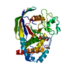
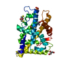
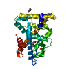
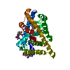
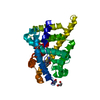
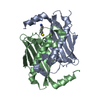
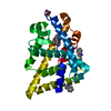
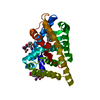
| Title | CRYSTAL STRUCTURE OF THE HUMAN PROTEIN TYROSINE PHOSPHATASE PTPN5 AT 1.8A RESOLUTION |
|---|
 Components Components | TYROSINE-PROTEIN PHOSPHATASE, NON-RECEPTOR TYPE 5 |
|---|
 Keywords Keywords | HYDROLASE / PTPN5 / STEP / PHOSPHATASE |
|---|
| Function / homology |  Function and homology information Function and homology information
Interleukin-37 signaling / protein dephosphorylation / phosphotyrosine residue binding / protein-tyrosine-phosphatase / protein tyrosine phosphatase activity / cell junction / protein kinase binding / endoplasmic reticulum membrane / signal transduction / nucleoplasm ...Interleukin-37 signaling / protein dephosphorylation / phosphotyrosine residue binding / protein-tyrosine-phosphatase / protein tyrosine phosphatase activity / cell junction / protein kinase binding / endoplasmic reticulum membrane / signal transduction / nucleoplasm / membrane / plasma membrane / cytosolSimilarity search - Function Protein-tyrosine phosphatase, receptor type R/non-receptor type 5 / Protein-tyrosine phosphatase, KIM-containing / Protein tyrosine phosphatase superfamily / Protein-Tyrosine Phosphatase; Chain A / Protein tyrosine phosphatase, catalytic domain / PTP type protein phosphatase domain profile. / Protein-tyrosine phosphatase / Tyrosine-specific protein phosphatase, PTPase domain / Protein-tyrosine phosphatase, catalytic / Protein tyrosine phosphatase, catalytic domain motif ...Protein-tyrosine phosphatase, receptor type R/non-receptor type 5 / Protein-tyrosine phosphatase, KIM-containing / Protein tyrosine phosphatase superfamily / Protein-Tyrosine Phosphatase; Chain A / Protein tyrosine phosphatase, catalytic domain / PTP type protein phosphatase domain profile. / Protein-tyrosine phosphatase / Tyrosine-specific protein phosphatase, PTPase domain / Protein-tyrosine phosphatase, catalytic / Protein tyrosine phosphatase, catalytic domain motif / Tyrosine specific protein phosphatases active site. / Protein-tyrosine phosphatase, active site / Tyrosine specific protein phosphatases domain profile. / Tyrosine-specific protein phosphatases domain / Protein-tyrosine phosphatase-like / Alpha-Beta Complex / Alpha BetaSimilarity search - Domain/homology |
|---|
| Biological species |  HOMO SAPIENS (human) HOMO SAPIENS (human) |
|---|
| Method |  X-RAY DIFFRACTION / SYNCHROTRON / MOLECULAR REPLACEMENT / Resolution: 1.8 Å X-RAY DIFFRACTION / SYNCHROTRON / MOLECULAR REPLACEMENT / Resolution: 1.8 Å |
|---|
 Authors Authors | Debreczeni, J.E. / Barr, A.J. / Eswaran, J. / Smee, C. / Burgess, N. / Gileadi, O. / von Delft, F. / Sundstrom, M. / Arrowsmith, C. / Edwards, A. / Knapp, S. |
|---|
 Citation Citation | Journal: Biochem. J. / Year: 2006
Title: Crystal structures and inhibitor identification for PTPN5, PTPRR and PTPN7: a family of human MAPK-specific protein tyrosine phosphatases.
Authors: Eswaran, J. / von Kries, J.P. / Marsden, B. / Longman, E. / Debreczeni, J.E. / Ugochukwu, E. / Turnbull, A. / Lee, W.H. / Knapp, S. / Barr, A.J. |
|---|
| History | | Deposition | Jun 22, 2005 | Deposition site: PDBE / Processing site: PDBE |
|---|
| Revision 1.0 | Jul 14, 2005 | Provider: repository / Type: Initial release |
|---|
| Revision 1.1 | Jul 13, 2011 | Group: Advisory / Version format compliance |
|---|
| Revision 1.2 | Jan 24, 2018 | Group: Structure summary / Category: audit_author / Item: _audit_author.name |
|---|
| Revision 1.3 | Feb 28, 2018 | Group: Database references / Source and taxonomy / Category: citation / citation_author / entity_src_gen
Item: _citation.journal_abbrev / _citation.journal_id_ISSN ..._citation.journal_abbrev / _citation.journal_id_ISSN / _citation.page_last / _citation.title / _citation_author.name / _entity_src_gen.pdbx_host_org_cell_line / _entity_src_gen.pdbx_host_org_ncbi_taxonomy_id / _entity_src_gen.pdbx_host_org_scientific_name / _entity_src_gen.pdbx_host_org_strain |
|---|
| Revision 1.4 | May 8, 2019 | Group: Data collection / Derived calculations / Experimental preparation
Category: exptl_crystal_grow / struct_conn
Item: _exptl_crystal_grow.method / _struct_conn.pdbx_leaving_atom_flag |
|---|
| Revision 1.5 | Oct 23, 2019 | Group: Data collection / Database references / Other / Category: pdbx_database_status / struct_ref_seq_dif
Item: _pdbx_database_status.status_code_sf / _struct_ref_seq_dif.details |
|---|
| Revision 1.6 | Dec 13, 2023 | Group: Data collection / Database references ...Data collection / Database references / Derived calculations / Refinement description
Category: chem_comp_atom / chem_comp_bond ...chem_comp_atom / chem_comp_bond / database_2 / pdbx_initial_refinement_model / struct_site
Item: _database_2.pdbx_DOI / _database_2.pdbx_database_accession ..._database_2.pdbx_DOI / _database_2.pdbx_database_accession / _struct_site.pdbx_auth_asym_id / _struct_site.pdbx_auth_comp_id / _struct_site.pdbx_auth_seq_id |
|---|
| Revision 1.7 | Oct 16, 2024 | Group: Structure summary / Category: pdbx_entry_details / pdbx_modification_feature / Item: _pdbx_entry_details.has_protein_modification |
|---|
|
|---|
 Movie
Movie Controller

 Yorodumi
 Open data
 Basic information
 Structure visualization
 Downloads & links
 Other downloads


 PDBj
PDBj


 Assembly


 Mass: 96.063 Da / Num. of mol.: 1 / Source method: obtained synthetically / Formula: SO4
 Mass: 92.094 Da / Num. of mol.: 1 / Source method: obtained synthetically / Formula: C3H8O3
 Mass: 18.015 Da / Num. of mol.: 271 / Source method: isolated from a natural source / Formula: H2O
 Sample preparation
 / Beamline: 14.1 / Wavelength: 0.9184
  Processing