 Movie
Movie Controller

[English] 日本語
 Yorodumi
- PDB-2bij: Crystal structure of the human protein tyrosine phosphatase PTPN5... -
+ Open data

- Basic information
| Entry | Database: PDB / ID: 2bij | ||||||
|---|---|---|---|---|---|---|---|
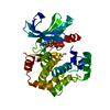
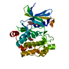
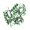
| Title | Crystal structure of the human protein tyrosine phosphatase PTPN5 (STEP, striatum enriched enriched Phosphatase) | ||||||
 Components Components | TYROSINE-PROTEIN PHOSPHATASE, NON-RECEPTOR TYPE 5 | ||||||
 Keywords Keywords | HYDROLASE / PTPN5 / STEP / PHOSPHATASE | ||||||
| Function / homology |  Function and homology information Function and homology informationInterleukin-37 signaling / protein dephosphorylation / phosphotyrosine residue binding / protein-tyrosine-phosphatase / protein tyrosine phosphatase activity / cell junction / protein kinase binding / endoplasmic reticulum membrane / signal transduction / nucleoplasm ...Interleukin-37 signaling / protein dephosphorylation / phosphotyrosine residue binding / protein-tyrosine-phosphatase / protein tyrosine phosphatase activity / cell junction / protein kinase binding / endoplasmic reticulum membrane / signal transduction / nucleoplasm / membrane / plasma membrane / cytosol Similarity search - Function | ||||||
| Biological species |  HOMO SAPIENS (human) HOMO SAPIENS (human) | ||||||
| Method |  X-RAY DIFFRACTION / MOLECULAR REPLACEMENT / Resolution: 2.05 Å X-RAY DIFFRACTION / MOLECULAR REPLACEMENT / Resolution: 2.05 Å | ||||||
 Authors Authors | Barr, A.J. / Debreczeni, J.E. / Eswaran, J. / Smee, C. / Burgess, N. / Gileadi, O. / Sundstrom, M. / Arrowsmith, C. / Edwards, A. / Knapp, S. / von Delft, F. | ||||||
 Citation Citation | Journal: Biochem. J. / Year: 2006 Title: Crystal structures and inhibitor identification for PTPN5, PTPRR and PTPN7: a family of human MAPK-specific protein tyrosine phosphatases. Authors: Eswaran, J. / von Kries, J.P. / Marsden, B. / Longman, E. / Debreczeni, J.E. / Ugochukwu, E. / Turnbull, A. / Lee, W.H. / Knapp, S. / Barr, A.J. | ||||||
| History |
|
- Structure visualization
| Structure viewer | Molecule: MolmilJmol/JSmol |
|---|
- Downloads & links
-Download
| PDBx/mmCIF format | 2bij.cif.gz | 73.3 KB | Display | PDBx/mmCIF format |
|---|---|---|---|---|
| PDB format | pdb2bij.ent.gz | 52.7 KB | Display | PDB format |
| PDBx/mmJSON format | 2bij.json.gz | Tree view | PDBx/mmJSON format | |
| Others |  Other downloads Other downloads |
-Validation report
| Arichive directory | https://data.pdbj.org/pub/pdb/validation_reports/bi/2bijftp://data.pdbj.org/pub/pdb/validation_reports/bi/2bij | HTTPS FTP |
|---|
-Related structure data
| Related structure data |  2a8bC 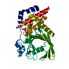 2bv5C  1jlnS S: Starting model for refinement C: citing same article ( |
|---|---|
| Similar structure data |


-Links
 PDBj
PDBj


- Assembly
| Deposited unit | 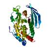
| ||||||||
|---|---|---|---|---|---|---|---|---|---|
| 1 |
| ||||||||
| Unit cell |
|
-Components
| #1: Protein | Mass: 35204.844 Da / Num. of mol.: 1 / Fragment: CATALYTIC DOMAIN, RESIDUES 258-539 Source method: isolated from a genetically manipulated source Source: (gene. exp.) HOMO SAPIENS (human) / Organ: BRAIN / Plasmid: PLIC SGC / Production host:  |
|---|---|
| #2: Chemical | ChemComp-SO4 /   Mass: 96.063 Da / Num. of mol.: 1 / Source method: obtained synthetically / Formula: SO4 Mass: 96.063 Da / Num. of mol.: 1 / Source method: obtained synthetically / Formula: SO4 |
| #3: Water | ChemComp-HOH /  Mass: 18.015 Da / Num. of mol.: 103 / Source method: isolated from a natural source / Formula: H2O Mass: 18.015 Da / Num. of mol.: 103 / Source method: isolated from a natural source / Formula: H2O |
| Sequence details | RESIDUES ASP 289, LEU 298, VAL 299 AND THR 517 ARE GIVEN AS VARIANTS IN THE SWISS-PROT ENTRY FOR ...RESIDUES ASP 289, LEU 298, VAL 299 AND THR 517 ARE GIVEN AS VARIANTS IN THE SWISS-PROT ENTRY FOR P54829. RESIDUES -23 TO -1 FORM PART OF A N-TERMINAL HIS-TAG USED FOR EXPRESSION |
-Experimental details
-Experiment
| Experiment | Method: X-RAY DIFFRACTION / Number of used crystals: 1 |
|---|
- Sample preparation
| Crystal | Density Matthews: 2.4 Å3/Da / Density % sol: 48 % |
|---|---|
| Crystal grow | Details: PROTEIN SOLUTION: 10 MG/ML PTPN5, 50 MM HEPES PH 7.5, 200 MM NACL, 10 MM DTT PRECIPITANT: 25% PEG3350, 0.2 M LI2SO4, 100 MM BIS-TR-S PH 5.5 |
-Data collection
| Diffraction | Mean temperature: 100 K |
|---|---|
| Diffraction source | Source: ROTATING ANODE / Type: RIGAKU / Wavelength: 1.5418 |
| Detector | Type: RIGAKU IMAGE PLATE / Detector: IMAGE PLATE / Date: Dec 2, 2004 / Details: MULTILAYER MIRRORS |
| Radiation | Protocol: SINGLE WAVELENGTH / Monochromatic (M) / Laue (L): M / Scattering type: x-ray |
| Radiation wavelength | Wavelength: 1.5418 Å / Relative weight: 1 |
| Reflection | Resolution: 2.05→39.74 Å / Num. obs: 20825 / % possible obs: 95 % / Observed criterion σ(I): 3 / Redundancy: 4.7 % / Rmerge(I) obs: 0.09 / Net I/σ(I): 11.14 |
| Reflection shell | Resolution: 2.05→2.15 Å / Redundancy: 3.9 % / Rmerge(I) obs: 0.37 / Mean I/σ(I) obs: 3.05 / % possible all: 97 |
- Processing
| Software |
| ||||||||||||||||||||||||||||||||||||||||||||||||||||||||||||||||||||||||||||||||||||||||||||||||||||||||||||||||||||||||||||||||||||||||||||||||||||||||||||||||||||||||||||||||||||||
|---|---|---|---|---|---|---|---|---|---|---|---|---|---|---|---|---|---|---|---|---|---|---|---|---|---|---|---|---|---|---|---|---|---|---|---|---|---|---|---|---|---|---|---|---|---|---|---|---|---|---|---|---|---|---|---|---|---|---|---|---|---|---|---|---|---|---|---|---|---|---|---|---|---|---|---|---|---|---|---|---|---|---|---|---|---|---|---|---|---|---|---|---|---|---|---|---|---|---|---|---|---|---|---|---|---|---|---|---|---|---|---|---|---|---|---|---|---|---|---|---|---|---|---|---|---|---|---|---|---|---|---|---|---|---|---|---|---|---|---|---|---|---|---|---|---|---|---|---|---|---|---|---|---|---|---|---|---|---|---|---|---|---|---|---|---|---|---|---|---|---|---|---|---|---|---|---|---|---|---|---|---|---|---|
| Refinement | Method to determine structure: MOLECULAR REPLACEMENT Starting model: PDB ENTRY 1JLN Resolution: 2.05→54.23 Å / Cor.coef. Fo:Fc: 0.938 / Cor.coef. Fo:Fc free: 0.895 / SU B: 9.811 / SU ML: 0.197 / TLS residual ADP flag: LIKELY RESIDUAL / Cross valid method: THROUGHOUT / ESU R: 0.22 / ESU R Free: 0.196 / Stereochemistry target values: MAXIMUM LIKELIHOOD Details: HYDROGENS HAVE BEEN ADDED IN THE RIDING POSITIONS. RESIDUES 380-382 ARE DISORDERED AND NOT VISIBLE IN THE ELECTRON DENSITY.
| ||||||||||||||||||||||||||||||||||||||||||||||||||||||||||||||||||||||||||||||||||||||||||||||||||||||||||||||||||||||||||||||||||||||||||||||||||||||||||||||||||||||||||||||||||||||
| Solvent computation | Ion probe radii: 0.8 Å / Shrinkage radii: 0.8 Å / VDW probe radii: 1.2 Å / Solvent model: BABINET MODEL WITH MASK | ||||||||||||||||||||||||||||||||||||||||||||||||||||||||||||||||||||||||||||||||||||||||||||||||||||||||||||||||||||||||||||||||||||||||||||||||||||||||||||||||||||||||||||||||||||||
| Displacement parameters | Biso mean: 36.21 Å2
| ||||||||||||||||||||||||||||||||||||||||||||||||||||||||||||||||||||||||||||||||||||||||||||||||||||||||||||||||||||||||||||||||||||||||||||||||||||||||||||||||||||||||||||||||||||||
| Refinement step | Cycle: LAST / Resolution: 2.05→54.23 Å
| ||||||||||||||||||||||||||||||||||||||||||||||||||||||||||||||||||||||||||||||||||||||||||||||||||||||||||||||||||||||||||||||||||||||||||||||||||||||||||||||||||||||||||||||||||||||
| Refine LS restraints |
|