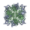
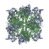
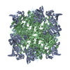
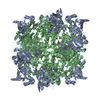
 Movie
Movie Controller

[English] 日本語
 Yorodumi
- PDB-2bv0: Crystal Structure of Protocatechuate 3,4-Dioxygenase from Acineto... -
+ Open data

- Basic information
| Entry | Database: PDB / ID: 2bv0 | ||||||
|---|---|---|---|---|---|---|---|
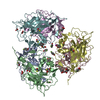
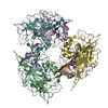
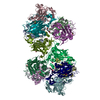
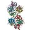
| Title | Crystal Structure of Protocatechuate 3,4-Dioxygenase from Acinetobacter Sp. ADP1 Mutant R133H in Complex with Protocatechuate. | ||||||
 Components Components |
| ||||||
 Keywords Keywords | OXIDOREDUCTASE / DIOXYGENASE / AROMATIC DEGRADATION / NON-HEME IRON / BETA-SANDWICH / MIXED ALPHA/BETA STRUCTURE | ||||||
| Function / homology |  Function and homology information Function and homology informationprotocatechuate 3,4-dioxygenase / protocatechuate 3,4-dioxygenase activity / 3,4-dihydroxybenzoate catabolic process / beta-ketoadipate pathway / ferric iron binding Similarity search - Function | ||||||
| Biological species |  ACINETOBACTER CALCOACETICUS (bacteria) ACINETOBACTER CALCOACETICUS (bacteria) | ||||||
| Method |  X-RAY DIFFRACTION / MOLECULAR REPLACEMENT / Resolution: 1.8 Å X-RAY DIFFRACTION / MOLECULAR REPLACEMENT / Resolution: 1.8 Å | ||||||
 Authors Authors | Vetting, M.W. / Valley, M.P. / D'Argenio, D.A. / Ornston, L.N. / Lipscomb, J.D. / Ohlendorf, D.H. | ||||||
 Citation Citation | Journal: Phd Thesis / Year: 2001 Title: Crystallographic Studies of Intradiol Dioxygenases. Authors: Vetting, M.W. | ||||||
| History |
| ||||||
| Remark 700 | SHEET THE SHEET STRUCTURE OF THIS MOLECULE IS BIFURCATED. IN ORDER TO REPRESENT THIS FEATURE IN ... SHEET THE SHEET STRUCTURE OF THIS MOLECULE IS BIFURCATED. IN ORDER TO REPRESENT THIS FEATURE IN THE SHEET RECORDS BELOW, TWO SHEETS ARE DEFINED. |
- Structure visualization
| Structure viewer | Molecule: MolmilJmol/JSmol |
|---|
- Downloads & links
-Download
| PDBx/mmCIF format | 2bv0.cif.gz | 109.2 KB | Display | PDBx/mmCIF format |
|---|---|---|---|---|
| PDB format | pdb2bv0.ent.gz | 82.1 KB | Display | PDB format |
| PDBx/mmJSON format | 2bv0.json.gz | Tree view | PDBx/mmJSON format | |
| Others |  Other downloads Other downloads |
-Validation report
| Summary document | 2bv0_validation.pdf.gz | 450.3 KB | Display | wwPDB validaton report |
|---|---|---|---|---|
| Full document | 2bv0_full_validation.pdf.gz | 456.3 KB | Display | |
| Data in XML | 2bv0_validation.xml.gz | 20.5 KB | Display | |
| Data in CIF | 2bv0_validation.cif.gz | 29.9 KB | Display | |
| Arichive directory | https://data.pdbj.org/pub/pdb/validation_reports/bv/2bv0ftp://data.pdbj.org/pub/pdb/validation_reports/bv/2bv0 | HTTPS FTP |
-Related structure data
| Related structure data |  2buuC  2buwC  2buxC  2buyC  1eo2S S: Starting model for refinement C: citing same article ( |
|---|---|
| Similar structure data |


-Links
 PDBj
PDBj- Assembly
| Deposited unit | 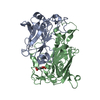
| ||||||||
|---|---|---|---|---|---|---|---|---|---|
| 1 | x 12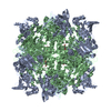
| ||||||||
| Unit cell |
| ||||||||
| Components on special symmetry positions |
| ||||||||
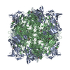
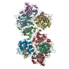
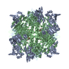
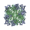
| Details | THE PHYSIOLOGICAL STATE OF THE MOLECULE IS A (AB)12DODECAMER. |
-Components
| #1: Protein | Mass: 23489.053 Da / Num. of mol.: 1 / Mutation: YES Source method: isolated from a genetically manipulated source Source: (gene. exp.) ACINETOBACTER CALCOACETICUS (bacteria) / Strain: ADP1 / Production host: References: UniProt: P20371, protocatechuate 3,4-dioxygenase |
|---|---|
| #2: Protein | Mass: 27583.031 Da / Num. of mol.: 1 Source method: isolated from a genetically manipulated source Source: (gene. exp.) ACINETOBACTER CALCOACETICUS (bacteria) / Strain: ADP1 / Production host: References: UniProt: P20372, protocatechuate 3,4-dioxygenase |
| #3: Chemical | ChemComp-FE /   Mass: 55.845 Da / Num. of mol.: 1 / Source method: obtained synthetically / Formula: Fe Mass: 55.845 Da / Num. of mol.: 1 / Source method: obtained synthetically / Formula: Fe |
| #4: Chemical | ChemComp-DHB / 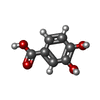  Mass: 154.120 Da / Num. of mol.: 1 / Source method: obtained synthetically / Formula: C7H6O4 Mass: 154.120 Da / Num. of mol.: 1 / Source method: obtained synthetically / Formula: C7H6O4 |
| #5: Water | ChemComp-HOH /  Mass: 18.015 Da / Num. of mol.: 242 / Source method: isolated from a natural source / Formula: H2O Mass: 18.015 Da / Num. of mol.: 242 / Source method: isolated from a natural source / Formula: H2O |
| Compound details | ENGINEERED |
-Experimental details
-Experiment
| Experiment | Method: X-RAY DIFFRACTION / Number of used crystals: 1 |
|---|
- Sample preparation
| Crystal | Density Matthews: 2.61 Å3/Da / Density % sol: 52.8 % Description: CRYSTAL WAS SOAKED IN 2.0 M AMMONIUM SULFATE, 100 MM TRIS PH 8.5, 30 MM PROTOCATECHUATE WITHIN AN ANEROBIC ENVIRONMENT PRIOR TO DATA COLLECTION. |
|---|---|
| Crystal grow | pH: 8.5 Details: 1.8 M AMMONIUM SULFATE, 100 MM TRIS-MALEATE PH 7.5, 0.08% PEG4000 PROTEIN AT 20 MG/ML |
-Data collection
| Diffraction | Mean temperature: 292 K |
|---|---|
| Diffraction source | Source: ROTATING ANODE / Type: RIGAKU RU200B / Wavelength: 1.5418 |
| Detector | Type: RIGAKU RAXIS IV / Detector: IMAGE PLATE / Details: OSMIC CONFOCAL MAXFLUX OPTICS |
| Radiation | Protocol: SINGLE WAVELENGTH / Monochromatic (M) / Laue (L): M / Scattering type: x-ray |
| Radiation wavelength | Wavelength: 1.5418 Å / Relative weight: 1 |
| Reflection | Resolution: 1.8→30 Å / Num. obs: 46726 / % possible obs: 99.5 % / Observed criterion σ(I): 0 / Redundancy: 3.4 % / Rmerge(I) obs: 0.04 / Net I/σ(I): 17.1 |
| Reflection shell | Resolution: 1.8→1.85 Å / Redundancy: 3.4 % / Rmerge(I) obs: 0.26 / Mean I/σ(I) obs: 5.8 / % possible all: 99.5 |
- Processing
| Software |
| ||||||||||||||||||||||||||||||||||||||||||||||||||||||||||||
|---|---|---|---|---|---|---|---|---|---|---|---|---|---|---|---|---|---|---|---|---|---|---|---|---|---|---|---|---|---|---|---|---|---|---|---|---|---|---|---|---|---|---|---|---|---|---|---|---|---|---|---|---|---|---|---|---|---|---|---|---|---|
| Refinement | Method to determine structure: MOLECULAR REPLACEMENT Starting model: PDB ENTRY 1EO2 Resolution: 1.8→30 Å / Cross valid method: THROUGHOUT / σ(F): 0
| ||||||||||||||||||||||||||||||||||||||||||||||||||||||||||||
| Displacement parameters | Biso mean: 26.8 Å2 | ||||||||||||||||||||||||||||||||||||||||||||||||||||||||||||
| Refinement step | Cycle: LAST / Resolution: 1.8→30 Å
| ||||||||||||||||||||||||||||||||||||||||||||||||||||||||||||
| Refine LS restraints |
| ||||||||||||||||||||||||||||||||||||||||||||||||||||||||||||
| LS refinement shell | Resolution: 1.8→1.86 Å /
|
