 Movie
Movie Controller

[English] 日本語
 Yorodumi
- PDB-2bum: Crystal Structure Of Wild-Type Protocatechuate 3,4-Dioxygenase fr... -
+ Open data

- Basic information
| Entry | Database: PDB / ID: 2bum | ||||||
|---|---|---|---|---|---|---|---|
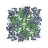
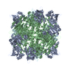
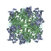
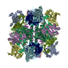
| Title | Crystal Structure Of Wild-Type Protocatechuate 3,4-Dioxygenase from Acinetobacter Sp. ADP1 | ||||||
 Components Components |
| ||||||
 Keywords Keywords | OXIDOREDUCTASE / AROMATIC DEGRADATION / NON-HEME IRON / BETA-SANDWICH / MIXED ALPHA/BETA STRUCTURE | ||||||
| Function / homology |  Function and homology information Function and homology informationprotocatechuate 3,4-dioxygenase / protocatechuate 3,4-dioxygenase activity / 3,4-dihydroxybenzoate catabolic process / beta-ketoadipate pathway / ferric iron binding Similarity search - Function | ||||||
| Biological species |  ACINETOBACTER SP. (bacteria) ACINETOBACTER SP. (bacteria) | ||||||
| Method |  X-RAY DIFFRACTION / MOLECULAR REPLACEMENT / Resolution: 1.8 Å X-RAY DIFFRACTION / MOLECULAR REPLACEMENT / Resolution: 1.8 Å | ||||||
 Authors Authors | Vetting, M.W. / Valley, M.P. / D'Argenio, D.A. / Ornston, L.N. / Lipscomb, J.D. / Ohlendorf, D.H. | ||||||
 Citation Citation | Journal: Annu.Rev.Microbiol. / Year: 2004 Title: Biophysical Analyses of Designed and Selected Mutants of Protocatechuate 3,4-Dioxygenase Authors: Brown, C.K. / Vetting, M.W. / Earhart, C.A. / Ohlendorf, D.H. #1: Journal: Ph D Thesis / Year: 2001Title: Crystallographic Studies of Intradiol Dioxygenases Authors: Vetting, M. | ||||||
| History |
| ||||||
| Remark 700 | SHEET THE SHEET STRUCTURE OF THIS MOLECULE IS BIFURCATED. IN ORDER TO REPRESENT THIS FEATURE IN ... SHEET THE SHEET STRUCTURE OF THIS MOLECULE IS BIFURCATED. IN ORDER TO REPRESENT THIS FEATURE IN THE SHEET RECORDS BELOW, TWO SHEETS ARE DEFINED. |
- Structure visualization
| Structure viewer | Molecule: MolmilJmol/JSmol |
|---|
- Downloads & links
-Download
| PDBx/mmCIF format | 2bum.cif.gz | 107.9 KB | Display | PDBx/mmCIF format |
|---|---|---|---|---|
| PDB format | pdb2bum.ent.gz | 81.1 KB | Display | PDB format |
| PDBx/mmJSON format | 2bum.json.gz | Tree view | PDBx/mmJSON format | |
| Others |  Other downloads Other downloads |
-Validation report
| Arichive directory | https://data.pdbj.org/pub/pdb/validation_reports/bu/2bumftp://data.pdbj.org/pub/pdb/validation_reports/bu/2bum | HTTPS FTP |
|---|
-Related structure data
| Related structure data |  2buqC  2burC  2butC  2buvC 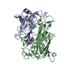 1eo2S S: Starting model for refinement C: citing same article ( |
|---|---|
| Similar structure data |


-Links
 PDBj
PDBj


- Assembly
| Deposited unit | 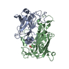
| ||||||||
|---|---|---|---|---|---|---|---|---|---|
| 1 | x 12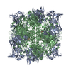
| ||||||||
| Unit cell |
| ||||||||
| Components on special symmetry positions |
| ||||||||
| Details | THE PHYSIOLOGICAL STATE OF THE MOLECULE IS A (AB)12DODECAMER.FOR THE HETERO-ASSEMBLY DESCRIBED BY REMARK 350 |
-Components
| #1: Protein | Mass: 23508.100 Da / Num. of mol.: 1 Source method: isolated from a genetically manipulated source Source: (gene. exp.) ACINETOBACTER SP. (bacteria) / Strain: ADP1 / Production host: References: UniProt: P20371, protocatechuate 3,4-dioxygenase |
|---|---|
| #2: Protein | Mass: 27583.031 Da / Num. of mol.: 1 Source method: isolated from a genetically manipulated source Source: (gene. exp.) ACINETOBACTER SP. (bacteria) / Strain: ADP1 / Production host: References: UniProt: P20372, protocatechuate 3,4-dioxygenase |
| #3: Chemical | ChemComp-FE /   Mass: 55.845 Da / Num. of mol.: 1 / Source method: obtained synthetically / Formula: Fe Mass: 55.845 Da / Num. of mol.: 1 / Source method: obtained synthetically / Formula: Fe |
| #4: Chemical | ChemComp-OH /   Mass: 17.007 Da / Num. of mol.: 1 / Source method: obtained synthetically / Formula: HO Mass: 17.007 Da / Num. of mol.: 1 / Source method: obtained synthetically / Formula: HO |
| #5: Water | ChemComp-HOH /  Mass: 18.015 Da / Num. of mol.: 209 / Source method: isolated from a natural source / Formula: H2O Mass: 18.015 Da / Num. of mol.: 209 / Source method: isolated from a natural source / Formula: H2O |
| Sequence details | RESIDUES ARE NUMBERED TO CORRELATE WITH RESIDUE NUMBERING OF 3,4-PCD FROM PSEUDOMONAS PUTIDA ...RESIDUES ARE NUMBERED TO CORRELATE WITH RESIDUE NUMBERING OF 3,4-PCD FROM PSEUDOMONA |
-Experimental details
-Experiment
| Experiment | Method: X-RAY DIFFRACTION / Number of used crystals: 1 |
|---|
- Sample preparation
| Crystal | Density Matthews: 2.61 Å3/Da / Density % sol: 52.3 % |
|---|---|
| Crystal grow | pH: 7.5 Details: 1.8 M AMMONIUM SULFATE, 100 MM TRIS-MALEATE PH 7.5, 0.08% PEG 4000, PROTEIN AT 20 MG/ML |
-Data collection
| Diffraction | Mean temperature: 194 K |
|---|---|
| Diffraction source | Source: ROTATING ANODE / Type: RIGAKU RU200B / Wavelength: 1.5418 |
| Detector | Type: RIGAKU R-AXIS IV / Details: OSMIC CONFOCAL MAXFLUX OPTICS |
| Radiation | Protocol: SINGLE WAVELENGTH / Monochromatic (M) / Laue (L): M / Scattering type: x-ray |
| Radiation wavelength | Wavelength: 1.5418 Å / Relative weight: 1 |
| Reflection | Resolution: 1.8→20 Å / Num. obs: 46943 / % possible obs: 99.5 % / Observed criterion σ(I): 0 / Redundancy: 3.5 % / Rmerge(I) obs: 0.06 / Net I/σ(I): 12 |
| Reflection shell | Resolution: 1.8→1.85 Å / Redundancy: 3.4 % / Rmerge(I) obs: 0.34 / Mean I/σ(I) obs: 4.4 / % possible all: 99.3 |
- Processing
| Software |
| ||||||||||||||||||||||||||||||||||||||||||||||||||||||||||||
|---|---|---|---|---|---|---|---|---|---|---|---|---|---|---|---|---|---|---|---|---|---|---|---|---|---|---|---|---|---|---|---|---|---|---|---|---|---|---|---|---|---|---|---|---|---|---|---|---|---|---|---|---|---|---|---|---|---|---|---|---|---|
| Refinement | Method to determine structure: MOLECULAR REPLACEMENT Starting model: PDB ENTRY 1EO2 Resolution: 1.8→20 Å / Cross valid method: THROUGHOUT / σ(F): 0
| ||||||||||||||||||||||||||||||||||||||||||||||||||||||||||||
| Refinement step | Cycle: LAST / Resolution: 1.8→20 Å
| ||||||||||||||||||||||||||||||||||||||||||||||||||||||||||||
| Refine LS restraints |
| ||||||||||||||||||||||||||||||||||||||||||||||||||||||||||||
| LS refinement shell | Resolution: 1.8→1.86 Å /
|
