 Movie
Movie Controller

[English] 日本語
 Yorodumi
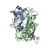
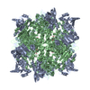
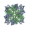
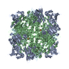
- PDB-1eoa: CRYSTAL STRUCTURE OF ACINETOBACTER SP. ADP1 PROTOCATECHUATE 3,4-D... -
+ Open data

- Basic information
| Entry | Database: PDB / ID: 1eoa | ||||||
|---|---|---|---|---|---|---|---|
| Title | CRYSTAL STRUCTURE OF ACINETOBACTER SP. ADP1 PROTOCATECHUATE 3,4-DIOXYGENASE IN COMPLEX WITH CYANIDE | ||||||
 Components Components |
| ||||||
 Keywords Keywords | OXIDOREDUCTASE / beta-sandwich mixed alpha/beta structure dioxygenase biodegradation | ||||||
| Function / homology |  Function and homology information Function and homology informationprotocatechuate 3,4-dioxygenase / protocatechuate 3,4-dioxygenase activity / 3,4-dihydroxybenzoate catabolic process / beta-ketoadipate pathway / ferric iron binding Similarity search - Function | ||||||
| Biological species |  Acinetobacter sp. (bacteria) Acinetobacter sp. (bacteria) | ||||||
| Method |  X-RAY DIFFRACTION / Resolution: 2.15 Å X-RAY DIFFRACTION / Resolution: 2.15 Å | ||||||
 Authors Authors | Vetting, M.W. / D'Argenio, D.A. / Ornston, L.N. / Ohlendorf, D.H. | ||||||
 Citation Citation | Journal: Biochemistry / Year: 2000 Title: Structure of Acinetobacter strain ADP1 protocatechuate 3, 4-dioxygenase at 2.2 A resolution: implications for the mechanism of an intradiol dioxygenase. Authors: Vetting, M.W. / D'Argenio, D.A. / Ornston, L.N. / Ohlendorf, D.H. #1: Journal: J.Mol.Biol. / Year: 1994Title: Crystallization and Preliminary X-ray Analysis of Protocatechuate 3,4-dioxygenase from Acinetobacter calcoaceticus Authors: Vetting, M.W. / Earhart, C.A. / Ohlendorf, D.H. #2: Journal: J.Bacteriol. / Year: 1999Title: Substitution, Insertion, Deletion, Suppression, and Altered Substrate Specificity in Functional Protocatechuate 3,4-dioxygenases. Authors: D'Argenio, D.A. / Vetting, M.W. / Ohlendorf, D.H. / Ornston, L.N. #3: Journal: Biochemistry / Year: 1997Title: Crystal Structures of Substrate and Substrate Analog Complexes of Protocatechuate 3,4-dioxygenase: Endogenous Fe3+ Ligand Displacement in Response to Substrate binding. Authors: Orville, A.M. / Lipscomb, J.D. / Ohlendorf, D.H. #4: Journal: J.Mol.Biol. / Year: 1994Title: Structure of Protocatechuate 3,4-dioxygenase from Pseudomonas aeruginosa at 2.15 A resolution Authors: Ohlendorf, D.H. / Orville, A.M. / Lipscomb, J.D. | ||||||
| History |
|
- Structure visualization
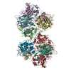
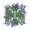
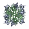
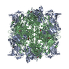
| Structure viewer | Molecule: MolmilJmol/JSmol |
|---|
- Downloads & links
-Download
| PDBx/mmCIF format | 1eoa.cif.gz | 105.8 KB | Display | PDBx/mmCIF format |
|---|---|---|---|---|
| PDB format | pdb1eoa.ent.gz | 80.8 KB | Display | PDB format |
| PDBx/mmJSON format | 1eoa.json.gz | Tree view | PDBx/mmJSON format | |
| Others |  Other downloads Other downloads |
-Validation report
| Arichive directory | https://data.pdbj.org/pub/pdb/validation_reports/eo/1eoaftp://data.pdbj.org/pub/pdb/validation_reports/eo/1eoa | HTTPS FTP |
|---|
-Related structure data


-Links
 PDBj
PDBj


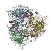
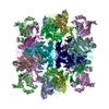
- Assembly
| Deposited unit | 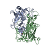
| ||||||||
|---|---|---|---|---|---|---|---|---|---|
| 1 | x 12
| ||||||||
| Unit cell |
| ||||||||
| Components on special symmetry positions |
| ||||||||
| Details | The biological assembly is a dodecamer (AB) X 12 constructed from the 23(T) symmetry of the space group acting on the A and B subunits in the assymetric unit. |
-Components
| #1: Protein | Mass: 23508.100 Da / Num. of mol.: 1 Source method: isolated from a genetically manipulated source Source: (gene. exp.) Acinetobacter sp. (bacteria) / Strain: ADP1 / Production host: References: UniProt: P20371, protocatechuate 3,4-dioxygenase | ||
|---|---|---|---|
| #2: Protein | Mass: 27583.031 Da / Num. of mol.: 1 Source method: isolated from a genetically manipulated source Source: (gene. exp.) Acinetobacter sp. (bacteria) / Strain: ADP1 / Production host: References: UniProt: P20372, protocatechuate 3,4-dioxygenase | ||
| #3: Chemical | ChemComp-FE /   Mass: 55.845 Da / Num. of mol.: 1 / Source method: obtained synthetically / Formula: Fe Mass: 55.845 Da / Num. of mol.: 1 / Source method: obtained synthetically / Formula: Fe | ||
| #4: Chemical |   Mass: 26.017 Da / Num. of mol.: 3 / Source method: obtained synthetically / Formula: CN Mass: 26.017 Da / Num. of mol.: 3 / Source method: obtained synthetically / Formula: CN#5: Water | ChemComp-HOH / |  Mass: 18.015 Da / Num. of mol.: 186 / Source method: isolated from a natural source / Formula: H2O Mass: 18.015 Da / Num. of mol.: 186 / Source method: isolated from a natural source / Formula: H2O |
-Experimental details
-Experiment
| Experiment | Method: X-RAY DIFFRACTION / Number of used crystals: 1 |
|---|
- Sample preparation
| Crystal | Density Matthews: 2.48 Å3/Da / Density % sol: 50.4 % | ||||||||||||||||||||
|---|---|---|---|---|---|---|---|---|---|---|---|---|---|---|---|---|---|---|---|---|---|
| Crystal grow | Temperature: 298 K / Method: vapor diffusion, hanging drop / pH: 7 Details: 100mM Tris-HCl pH 7.0, 2.0 M ammonium sulfate, VAPOR DIFFUSION, HANGING DROP, temperature 298K Crystals were soaked in 200mM NaCN at pH 8.5 | ||||||||||||||||||||
| Crystal grow | *PLUS Temperature: 18 ℃ | ||||||||||||||||||||
| Components of the solutions | *PLUS
|
-Data collection
| Diffraction | Mean temperature: 298 K |
|---|---|
| Diffraction source | Source: ROTATING ANODE / Type: RIGAKU RU200 / Wavelength: 1.5418 |
| Detector | Type: SIEMENS HI-STAR / Detector: AREA DETECTOR / Date: Jan 1, 1999 |
| Radiation | Protocol: SINGLE WAVELENGTH / Monochromatic (M) / Laue (L): M / Scattering type: x-ray |
| Radiation wavelength | Wavelength: 1.5418 Å / Relative weight: 1 |
| Reflection | Resolution: 2.15→20 Å / Num. all: 126684 / Num. obs: 27540 / % possible obs: 98.2 % / Observed criterion σ(F): 0 / Observed criterion σ(I): 1 / Redundancy: 4.6 % / Rmerge(I) obs: 0.087 / Net I/σ(I): 8.7 |
| Reflection shell | Resolution: 2.15→2.2 Å / Redundancy: 2.2 % / Rmerge(I) obs: 0.305 / % possible all: 90.6 |
| Reflection shell | *PLUS % possible obs: 90.6 % / Mean I/σ(I) obs: 1 |
- Processing
| Software |
| |||||||||||||||||||||||||
|---|---|---|---|---|---|---|---|---|---|---|---|---|---|---|---|---|---|---|---|---|---|---|---|---|---|---|
| Refinement | Resolution: 2.15→20 Å / σ(F): 0 / σ(I): 0 / Stereochemistry target values: Engh & Huber
| |||||||||||||||||||||||||
| Refinement step | Cycle: LAST / Resolution: 2.15→20 Å
| |||||||||||||||||||||||||
| Refine LS restraints |
| |||||||||||||||||||||||||
| Software | *PLUS Name: 'CNS' / Classification: refinement | |||||||||||||||||||||||||
| Refine LS restraints | *PLUS Type: c_bond_d / Dev ideal: 0.006 |
