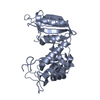
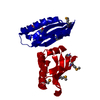
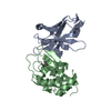
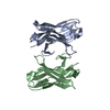
SHEET THE SHEET STRUCTURE OF THIS MOLECULE IS BIFURCATED. IN ORDER TO REPRESENT THIS FEATURE IN ... SHEET THE SHEET STRUCTURE OF THIS MOLECULE IS BIFURCATED. IN ORDER TO REPRESENT THIS FEATURE IN THE SHEET RECORDS BELOW, TWO SHEETS ARE DEFINED.
Mass: 18.015 Da / Num. of mol.: 50 / Source method: isolated from a natural source / Formula: H2O
Nonpolymer details
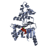
GLUTATHIONE (GTT): GLUTATHIONES X1 AND X2 ARE COVALENTLY ATTACHED TO A 2293 CYS AND B 2293 CYS, ...GLUTATHIONE (GTT): GLUTATHIONES X1 AND X2 ARE COVALENTLY ATTACHED TO A 2293 CYS AND B 2293 CYS, RESPECTIVELY, FORMING A DISULFIDE BOND.
Sequence details
THE FIRST THREE RESIDUES GAM ORIGINATES FROM THE EXPRESSION PLASMID.
-
Experimental details
-
Experiment
Experiment
Method: X-RAY DIFFRACTION / Number of used crystals: 1
-
Sample preparation
Crystal
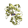
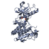
Density Matthews: 2.6 Å3/Da / Density % sol: 52.6 % / Description: FILAMIN C REPEAT 24
Crystal grow
pH: 5.5 / Details: 1.26 M SODIUM CITRATE, 0.1 M CITRIC ACID PH 5.5
Resolution: 2.1→37.68 Å / Cor.coef. Fo:Fc: 0.954 / Cor.coef. Fo:Fc free: 0.925 / SU B: 10.227 / SU ML: 0.136 / TLS residual ADP flag: LIKELY RESIDUAL / Cross valid method: THROUGHOUT / ESU R: 0.228 / ESU R Free: 0.196 / Stereochemistry target values: MAXIMUM LIKELIHOOD Details: HYDROGENS HAVE BEEN ADDED IN THE RIDING POSITIONS. SOME OF THE SIDE CHAIN ATOMS OF RESIDUES A 2264 ARG, A 2280 LYS, A 2287 ASP, B 2240 LYS, B 2250 ARG, B 2262 TRP, B 2280 LYS, B 2287 ASP, B ...Details: HYDROGENS HAVE BEEN ADDED IN THE RIDING POSITIONS. SOME OF THE SIDE CHAIN ATOMS OF RESIDUES A 2264 ARG, A 2280 LYS, A 2287 ASP, B 2240 LYS, B 2250 ARG, B 2262 TRP, B 2280 LYS, B 2287 ASP, B 2288 ARG, D 785 THR AND D 787 ASN HAVE A POORLY DEFINED DENSITY. THE SIDE CHAIN ATOMS OF RESIDUES A 2289 LYS, B 2239 HIS, B 2264 ARG, B 2286 GLU, B 2289 LYS AND B 2314 GLU HAVE NO ELECTRON DENSITY BUT THEY WERE MODELED. BREAK IN THE MAIN CHAIN ELECTRON DENSITY BETWEEN RESIDUES B 2286 GLU - B 2287 ASP, B 2288 ARG - B 2289 LYS AND D 786 ILE - D 787 ASN.
Rfactor
Num. reflection
% reflection
Selection details
Rfree
0.243
1381
10 %
RANDOM
Rwork
0.192
-
-
-
obs
0.197
12367
97.2 %
-
Solvent computation
Ion probe radii: 0.8 Å / Shrinkage radii: 0.8 Å / VDW probe radii: 1.2 Å / Solvent model: MASK
 Movie
Movie Controller

 Yorodumi
 Open data
 Basic information
 Components
 Keywords
 Function and homology information
 HOMO SAPIENS (human)
 X-RAY DIFFRACTION /
  Authors
 Citation
 Structure visualization
 Downloads & links
 Other downloads


 PDBj
PDBj


 Assembly
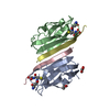


 Mass: 307.323 Da / Num. of mol.: 2 / Source method: obtained synthetically / Formula: C10H17N3O6S
 Mass: 92.094 Da / Num. of mol.: 2 / Source method: obtained synthetically / Formula: C3H8O3
 Mass: 18.015 Da / Num. of mol.: 50 / Source method: isolated from a natural source / Formula: H2O
 Sample preparation
 / Beamline: ID14-3 / Wavelength: 0.931
  Processing