 Movie
Movie Controller

[English] 日本語
 Yorodumi
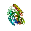
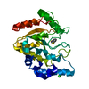
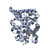
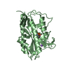
- PDB-2boo: The crystal structure of Uracil-DNA N-Glycosylase (UNG) from Dein... -
+ Open data

- Basic information
| Entry | Database: PDB / ID: 2boo | ||||||
|---|---|---|---|---|---|---|---|
| Title | The crystal structure of Uracil-DNA N-Glycosylase (UNG) from Deinococcus radiodurans. | ||||||
 Components Components | URACIL-DNA GLYCOSYLASE | ||||||
 Keywords Keywords | HYDROLASE / BASE EXCISION REPAIR / RADIATION RESISTANCE / DNA DAMAGE / DNA REPAIR / GLYCOSIDASE | ||||||
| Function / homology |  Function and homology information Function and homology informationbase-excision repair, AP site formation via deaminated base removal / uracil-DNA glycosylase / uracil DNA N-glycosylase activity / cytoplasm Similarity search - Function | ||||||
| Biological species |  DEINOCOCCUS RADIODURANS (radioresistant) DEINOCOCCUS RADIODURANS (radioresistant) | ||||||
| Method |  X-RAY DIFFRACTION / SYNCHROTRON / MOLECULAR REPLACEMENT / Resolution: 1.8 Å X-RAY DIFFRACTION / SYNCHROTRON / MOLECULAR REPLACEMENT / Resolution: 1.8 Å | ||||||
 Authors Authors | Leiros, I. / Moe, E. / Smalas, A.O. / McSweeney, S. | ||||||
 Citation Citation | Journal: Acta Crystallogr.,Sect.D / Year: 2005 Title: Structure of the Uracil-DNA N-Glycosylase (Ung) from Deinococcus Radiodurans. Authors: Leiros, I. / Moe, E. / Smalas, A.O. / Mcsweeney, S. | ||||||
| History |
|
- Structure visualization
| Structure viewer | Molecule: MolmilJmol/JSmol |
|---|
- Downloads & links
-Download
| PDBx/mmCIF format | 2boo.cif.gz | 63.4 KB | Display | PDBx/mmCIF format |
|---|---|---|---|---|
| PDB format | pdb2boo.ent.gz | 46 KB | Display | PDB format |
| PDBx/mmJSON format | 2boo.json.gz | Tree view | PDBx/mmJSON format | |
| Others |  Other downloads Other downloads |
-Validation report
| Arichive directory | https://data.pdbj.org/pub/pdb/validation_reports/bo/2booftp://data.pdbj.org/pub/pdb/validation_reports/bo/2boo | HTTPS FTP |
|---|
-Related structure data
| Related structure data | 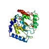 1akzS S: Starting model for refinement |
|---|---|
| Similar structure data |


-Links
 PDBj
PDBj- Assembly
| Deposited unit | 
| ||||||||
|---|---|---|---|---|---|---|---|---|---|
| 1 |
| ||||||||
| Unit cell |
|
-Components
| #1: Protein | Mass: 27780.500 Da / Num. of mol.: 1 Source method: isolated from a genetically manipulated source Details: RESIDUES 1-16 AND 247 NOT VISIBLE IN ELECTRON DENSITY Source: (gene. exp.) DEINOCOCCUS RADIODURANS (radioresistant)Strain: R1 / Production host: | ||||
|---|---|---|---|---|---|
| #2: Chemical |   Mass: 62.005 Da / Num. of mol.: 3 / Source method: obtained synthetically / Formula: NO3 Mass: 62.005 Da / Num. of mol.: 3 / Source method: obtained synthetically / Formula: NO3#3: Water | ChemComp-HOH / |  Mass: 18.015 Da / Num. of mol.: 154 / Source method: isolated from a natural source / Formula: H2O Mass: 18.015 Da / Num. of mol.: 154 / Source method: isolated from a natural source / Formula: H2OCompound details | EXCISES URACIL RESIDUES FROM THE DNA | |
-Experimental details
-Experiment
| Experiment | Method: X-RAY DIFFRACTION / Number of used crystals: 1 |
|---|
- Sample preparation
| Crystal | Density Matthews: 4.1 Å3/Da / Density % sol: 70 % |
|---|---|
| Crystal grow | Temperature: 277 K / pH: 7.5 Details: 0.2 M AMMONIUM NITRATE, 17% (W/V) PEG 3000 AT 4 DEGREES C, pH 7.50 |
-Data collection
| Diffraction | Mean temperature: 100 K |
|---|---|
| Diffraction source | Source: SYNCHROTRON / Site: ESRF  / Beamline: ID23-1 / Wavelength: 0.976 / Beamline: ID23-1 / Wavelength: 0.976 |
| Detector | Type: MARRESEARCH / Detector: CCD / Date: Dec 3, 2003 / Details: TOROIDAL MIRROR |
| Radiation | Monochromator: SILICON CRYSTAL / Protocol: SINGLE WAVELENGTH / Monochromatic (M) / Laue (L): M / Scattering type: x-ray |
| Radiation wavelength | Wavelength: 0.976 Å / Relative weight: 1 |
| Reflection | Resolution: 1.8→28.36 Å / Num. obs: 40425 / % possible obs: 93.7 % / Observed criterion σ(I): 0 / Redundancy: 2.52 % / Biso Wilson estimate: 20 Å2 / Rmerge(I) obs: 0.1 / Net I/σ(I): 4.44 |
| Reflection shell | Resolution: 1.8→1.9 Å / Redundancy: 2.37 % / Rmerge(I) obs: 0.27 / Mean I/σ(I) obs: 2.65 / % possible all: 95.2 |
- Processing
| Software |
| ||||||||||||||||||||||||||||||||||||||||||||||||||||||||||||||||||||||||||||||||||||||||||||||||||||||||||||||||||||||||||||||||||||||||||||||||||||||||||||||||||||||||||||||||||||||
|---|---|---|---|---|---|---|---|---|---|---|---|---|---|---|---|---|---|---|---|---|---|---|---|---|---|---|---|---|---|---|---|---|---|---|---|---|---|---|---|---|---|---|---|---|---|---|---|---|---|---|---|---|---|---|---|---|---|---|---|---|---|---|---|---|---|---|---|---|---|---|---|---|---|---|---|---|---|---|---|---|---|---|---|---|---|---|---|---|---|---|---|---|---|---|---|---|---|---|---|---|---|---|---|---|---|---|---|---|---|---|---|---|---|---|---|---|---|---|---|---|---|---|---|---|---|---|---|---|---|---|---|---|---|---|---|---|---|---|---|---|---|---|---|---|---|---|---|---|---|---|---|---|---|---|---|---|---|---|---|---|---|---|---|---|---|---|---|---|---|---|---|---|---|---|---|---|---|---|---|---|---|---|---|
| Refinement | Method to determine structure: MOLECULAR REPLACEMENT Starting model: PDB ENTRY 1AKZ Resolution: 1.8→84.52 Å / Cor.coef. Fo:Fc: 0.929 / Cor.coef. Fo:Fc free: 0.903 / SU B: 9.074 / SU ML: 0.119 / TLS residual ADP flag: LIKELY RESIDUAL / Cross valid method: THROUGHOUT / ESU R: 0.114 / ESU R Free: 0.117 / Stereochemistry target values: MAXIMUM LIKELIHOOD Details: HYDROGENS HAVE BEEN ADDED IN THE RIDING POSITIONS. SUB-OPTIMAL REFINEMENT STATISTICS CAUSED BY CRYSTALS CONSISTING OF SEVERAL LATTICES. FLEXIBLE REGIONS IN PROTEIN CAN NOT BE MODELLED.
| ||||||||||||||||||||||||||||||||||||||||||||||||||||||||||||||||||||||||||||||||||||||||||||||||||||||||||||||||||||||||||||||||||||||||||||||||||||||||||||||||||||||||||||||||||||||
| Solvent computation | Ion probe radii: 0.8 Å / Shrinkage radii: 0.8 Å / VDW probe radii: 1.2 Å / Solvent model: BABINET MODEL WITH MASK | ||||||||||||||||||||||||||||||||||||||||||||||||||||||||||||||||||||||||||||||||||||||||||||||||||||||||||||||||||||||||||||||||||||||||||||||||||||||||||||||||||||||||||||||||||||||
| Displacement parameters | Biso mean: 23.19 Å2
| ||||||||||||||||||||||||||||||||||||||||||||||||||||||||||||||||||||||||||||||||||||||||||||||||||||||||||||||||||||||||||||||||||||||||||||||||||||||||||||||||||||||||||||||||||||||
| Refinement step | Cycle: LAST / Resolution: 1.8→84.52 Å
| ||||||||||||||||||||||||||||||||||||||||||||||||||||||||||||||||||||||||||||||||||||||||||||||||||||||||||||||||||||||||||||||||||||||||||||||||||||||||||||||||||||||||||||||||||||||
| Refine LS restraints |
|