- PDB-2bmd: high resolution structure of GDP-bound human Rab4a -
+
Open data
ID or keywords:
Loading...
-
Basic information
Entry
Database: PDB / ID: 2bmd
Title
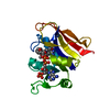
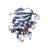
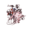
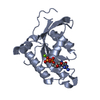
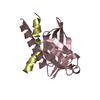
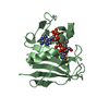
high resolution structure of GDP-bound human Rab4a
Components
RAS-RELATED PROTEIN RAB4A
Keywords
PROTEIN TRANSPORT / GTP-BINDING PROTEIN / VESICULAR TRANSPORT / ENDOCYTOSIS / PRENYLATION / TRANSPORT
Function / homology
Function and homology information
neurotransmitter receptor transport postsynaptic membrane to endosome / Rab protein signal transduction / insulin-responsive compartment / vesicle-mediated transport in synapse / RAB geranylgeranylation / MET receptor recycling / TBC/RABGAPs / antigen processing and presentation / Synthesis of PIPs at the plasma membrane / regulation of endocytosis ...neurotransmitter receptor transport postsynaptic membrane to endosome / Rab protein signal transduction / insulin-responsive compartment / vesicle-mediated transport in synapse / RAB geranylgeranylation / MET receptor recycling / TBC/RABGAPs / antigen processing and presentation / Synthesis of PIPs at the plasma membrane / regulation of endocytosis / vesicle-mediated transport / cytoplasmic vesicle membrane / small monomeric GTPase / Translocation of SLC2A4 (GLUT4) to the plasma membrane / recycling endosome / recycling endosome membrane / GDP binding / protein transport / G protein activity / early endosome membrane / vesicle / GTPase activity / GTP binding / perinuclear region of cytoplasm / glutamatergic synapse / extracellular exosome / metal ion binding Similarity search - Function
Rab4 / : / ARF-like small GTPases; ARF, ADP-ribosylation factor / Small GTPase Rab domain profile. / Ran (Ras-related nuclear proteins) /TC4 subfamily of small GTPases / Rho (Ras homology) subfamily of Ras-like small GTPases / Ras subfamily of RAS small GTPases / Small GTPase / Ras family / Rab subfamily of small GTPases ...Rab4 / : / ARF-like small GTPases; ARF, ADP-ribosylation factor / Small GTPase Rab domain profile. / Ran (Ras-related nuclear proteins) /TC4 subfamily of small GTPases / Rho (Ras homology) subfamily of Ras-like small GTPases / Ras subfamily of RAS small GTPases / Small GTPase / Ras family / Rab subfamily of small GTPases / Small GTP-binding protein domain / P-loop containing nucleotide triphosphate hydrolases / Rossmann fold / P-loop containing nucleoside triphosphate hydrolase / 3-Layer(aba) Sandwich / Alpha Beta Similarity search - Domain/homology
Mass: 18.015 Da / Num. of mol.: 208 / Source method: isolated from a natural source / Formula: H2O
-
Experimental details
-
Experiment
Experiment
Method: X-RAY DIFFRACTION / Number of used crystals: 1
-
Sample preparation
Crystal
Density Matthews: 1.79 Å3/Da / Density % sol: 30.68 % Description: PARTIALLY REFINED GPPNHP-BOUND RAB4A STRUCTURE WAS USED FOR MOLECULAR REPLACEMENT, SOLVED BY SAME AUTHORS
Crystal grow
Temperature: 293 K / Method: vapor diffusion, hanging drop Details: 30% (W/V) PEG 8000, 200MM AMMONIUM SULPHATE, 100MM SODIUM CACODYLATE PH6.5 HANGING DROP 20 DEGREE CELSIUS
Monochromator: MIRROR / Protocol: SINGLE WAVELENGTH / Monochromatic (M) / Laue (L): M / Scattering type: x-ray
Radiation wavelength
Wavelength: 1.0721 Å / Relative weight: 1
Reflection
Resolution: 1.8→20 Å / Num. obs: 15669 / % possible obs: 99.8 % / Redundancy: 4.3 % / Rmerge(I) obs: 0.07 / Net I/σ(I): 14.44
Reflection shell
Resolution: 1.8→1.86 Å / Rmerge(I) obs: 0.2 / Mean I/σ(I) obs: 7.5 / % possible all: 100
-
Processing
Software
Name
Version
Classification
REFMAC
5.1.19
refinement
XDS
datareduction
XSCALE
datascaling
MOLREP
phasing
Refinement
Method to determine structure: MOLECULAR REPLACEMENT / Resolution: 1.8→18.29 Å / Cor.coef. Fo:Fc: 0.958 / Cor.coef. Fo:Fc free: 0.933 / SU B: 2.388 / SU ML: 0.076 / Cross valid method: THROUGHOUT / ESU R: 0.13 / ESU R Free: 0.124 / Stereochemistry target values: MAXIMUM LIKELIHOOD Details: HYDROGENS HAVE BEEN ADDED IN THE RIDING POSITIONS. DISORDERED REGIONS WERE MODELED STEREOCHEMICALLY
Rfactor
Num. reflection
% reflection
Selection details
Rfree
0.205
783
5 %
RANDOM
Rwork
0.16
-
-
-
obs
0.162
14883
99.9 %
-
Solvent computation
Ion probe radii: 0.8 Å / Shrinkage radii: 0.8 Å / VDW probe radii: 1.4 Å / Solvent model: BABINET MODEL WITH MASK
 Movie
Movie Controller


 Open data
 Basic information
 Components
 Keywords
 Function and homology information
 HOMO SAPIENS (human)
 X-RAY DIFFRACTION /
  Authors
 Citation
 Structure visualization
 Downloads & links
 Other downloads

 PDBj
PDBj


 Assembly
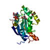


 Type: RNA linking / Mass: 443.201 Da / Num. of mol.: 1 / Source method: obtained synthetically / Formula: C10H15N5O11P2 / Comment: GDP, energy-carrying molecule*YM
 Mass: 92.094 Da / Num. of mol.: 2 / Source method: obtained synthetically / Formula: C3H8O3
 Mass: 18.015 Da / Num. of mol.: 208 / Source method: isolated from a natural source / Formula: H2O
 Sample preparation
 / Beamline: 14.1 / Wavelength: 1.0721
  Processing