regulation of cellular response to stress / DNA-binding transcription activator activity / phosphorelay signal transduction system / cis-regulatory region sequence-specific DNA binding / protein-DNA complex / transcription regulator complex / sequence-specific DNA binding / negative regulation of DNA-templated transcription / regulation of DNA-templated transcription / positive regulation of DNA-templated transcription ...regulation of cellular response to stress / DNA-binding transcription activator activity / phosphorelay signal transduction system / cis-regulatory region sequence-specific DNA binding / protein-DNA complex / transcription regulator complex / sequence-specific DNA binding / negative regulation of DNA-templated transcription / regulation of DNA-templated transcription / positive regulation of DNA-templated transcription / DNA binding / ATP binding / identical protein binding / cytoplasm Similarity search - Function
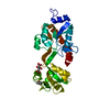
Journal: Science / Year: 2005 Title: Structural insights into the activity of enhancer-binding proteins. Authors: Mathieu Rappas / Jorg Schumacher / Fabienne Beuron / Hajime Niwa / Patricia Bordes / Sivaramesh Wigneshweraraj / Catherine A Keetch / Carol V Robinson / Martin Buck / Xiaodong Zhang / Abstract: Activators of bacterial sigma54-RNA polymerase holoenzyme are mechanochemical proteins that use adenosine triphosphate (ATP) hydrolysis to activate transcription. We have determined by cryogenic ...Activators of bacterial sigma54-RNA polymerase holoenzyme are mechanochemical proteins that use adenosine triphosphate (ATP) hydrolysis to activate transcription. We have determined by cryogenic electron microscopy (cryo-EM) a 20 angstrom resolution structure of an activator, phage shock protein F [PspF(1-275)], which is bound to an ATP transition state analog in complex with its basal factor, sigma54. By fitting the crystal structure of PspF(1-275) at 1.75 angstroms into the EM map, we identified two loops involved in binding sigma54. Comparing enhancer-binding structures in different nucleotide states and mutational analysis led us to propose nucleotide-dependent conformational changes that free the loops for association with sigma54.
Resolution: 1.7→37 Å / Num. obs: 29411 / % possible obs: 91.6 % / Observed criterion σ(I): 2.5 / Redundancy: 3.1 % / Rmerge(I) obs: 0.044 / Net I/σ(I): 26
Reflection shell
Resolution: 1.7→1.76 Å / Rmerge(I) obs: 0.277 / Mean I/σ(I) obs: 3 / % possible all: 60.5
-
Processing
Software
Name
Version
Classification
CNS
1.1
refinement
DENZO
datareduction
SCALEPACK
datascaling
SHARP
phasing
Refinement
Method to determine structure: MAD / Resolution: 1.7→37 Å / Data cutoff high absF: 100000000 / Isotropic thermal model: RESTRAINED / Cross valid method: THROUGHOUT / Stereochemistry target values: MAXIMUM LIKELIHOOD Details: HYDROGENS HAVE BEEN ADDED IN THE RIDING POSITIONS. DISORDERED REGIONS WERE MODELLED AS ALANINES
Rfactor
Num. reflection
% reflection
Selection details
Rfree
0.21244
2004
6.9 %
RANDOM
Rwork
0.17235
-
-
-
obs
0.17235
26845
91.6 %
-
Solvent computation
Solvent model: BABINET MODEL WITH MASK / Bsol: 300 Å2 / ksol: 0.8 e/Å3
Displacement parameters
Biso mean: 22.879 Å2
Baniso -1
Baniso -2
Baniso -3
1-
-1.11 Å2
-0.56 Å2
0 Å2
2-
-
-1.11 Å2
0 Å2
3-
-
-
1.67 Å2
Refinement step
Cycle: LAST / Resolution: 1.7→37 Å
Protein
Nucleic acid
Ligand
Solvent
Total
Num. atoms
1824
0
0
162
1986
Refine LS restraints
Refine-ID
Type
Dev ideal
Dev ideal target
X-RAY DIFFRACTION
c_bond_d
0.019
X-RAY DIFFRACTION
c_bond_d_na
X-RAY DIFFRACTION
c_bond_d_prot
X-RAY DIFFRACTION
c_angle_d
X-RAY DIFFRACTION
c_angle_d_na
X-RAY DIFFRACTION
c_angle_d_prot
X-RAY DIFFRACTION
c_angle_deg
1.6
X-RAY DIFFRACTION
c_angle_deg_na
X-RAY DIFFRACTION
c_angle_deg_prot
X-RAY DIFFRACTION
c_dihedral_angle_d
5.38
X-RAY DIFFRACTION
c_dihedral_angle_d_na
X-RAY DIFFRACTION
c_dihedral_angle_d_prot
X-RAY DIFFRACTION
c_improper_angle_d
X-RAY DIFFRACTION
c_improper_angle_d_na
X-RAY DIFFRACTION
c_improper_angle_d_prot
X-RAY DIFFRACTION
c_mcbond_it
1.236
1.5
X-RAY DIFFRACTION
c_mcangle_it
2.229
2
X-RAY DIFFRACTION
c_scbond_it
3.33
3
X-RAY DIFFRACTION
c_scangle_it
5.86
4.5
LS refinement shell
Resolution: 1.7→1.75 Å / Total num. of bins used: 20
In the structure databanks used in Yorodumi, some data are registered as the other names, "COVID-19 virus" and "2019-nCoV". Here are the details of the virus and the list of structure data.
Jan 31, 2019. EMDB accession codes are about to change! (news from PDBe EMDB page)
EMDB accession codes are about to change! (news from PDBe EMDB page)
The allocation of 4 digits for EMDB accession codes will soon come to an end. Whilst these codes will remain in use, new EMDB accession codes will include an additional digit and will expand incrementally as the available range of codes is exhausted. The current 4-digit format prefixed with “EMD-” (i.e. EMD-XXXX) will advance to a 5-digit format (i.e. EMD-XXXXX), and so on. It is currently estimated that the 4-digit codes will be depleted around Spring 2019, at which point the 5-digit format will come into force.
The EM Navigator/Yorodumi systems omit the EMD- prefix.
Related info.:Q: What is EMD? / ID/Accession-code notation in Yorodumi/EM Navigator
Yorodumi is a browser for structure data from EMDB, PDB, SASBDB, etc.
This page is also the successor to EM Navigator detail page, and also detail information page/front-end page for Omokage search.
The word "yorodu" (or yorozu) is an old Japanese word meaning "ten thousand". "mi" (miru) is to see.
Related info.:EMDB / PDB / SASBDB / Comparison of 3 databanks / Yorodumi Search / Aug 31, 2016. New EM Navigator & Yorodumi / Yorodumi Papers / Jmol/JSmol / Function and homology information / Changes in new EM Navigator and Yorodumi
 Movie
Movie Controller


 Open data
 Basic information
 Components
 Keywords
 Function and homology information
 X-RAY DIFFRACTION /
  Authors
 Citation
 Structure visualization
 Downloads & links
 Other downloads
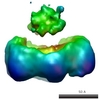


 PDBj
PDBj Assembly
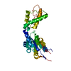
 Mass: 18.015 Da / Num. of mol.: 162 / Source method: isolated from a natural source / Formula: H2O
 Sample preparation
 / Beamline: ID29 / Wavelength: 0.97, 1.1
 Processing