 Movie
Movie Controller

[English] 日本語
 Yorodumi
- PDB-2c96: Structural basis of the nucleotide driven conformational changes ... -
+ Open data

- Basic information
| Entry | Database: PDB / ID: 2c96 | ||||||
|---|---|---|---|---|---|---|---|
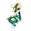
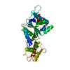
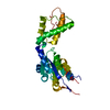
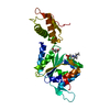
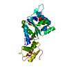
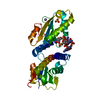
| Title | Structural basis of the nucleotide driven conformational changes in the AAA domain of transcription activator PspF | ||||||
 Components Components | PSP OPERON TRANSCRIPTIONAL ACTIVATOR | ||||||
 Keywords Keywords | TRANSCRIPTION REGULATION / BACTERIAL SIGMA54 ACTIVATOR / ATPASE / AAA DOMAIN / ACTIVATOR / ATP-BINDING / DNA-BINDING / NUCLEOTIDE-BINDING / SENSORY TRANSDUCTION / TWO-COMPONENT REGULATORY SYSTEM | ||||||
| Function / homology |  Function and homology information Function and homology informationregulation of cellular response to stress / DNA-binding transcription activator activity / phosphorelay signal transduction system / cis-regulatory region sequence-specific DNA binding / protein-DNA complex / transcription regulator complex / sequence-specific DNA binding / negative regulation of DNA-templated transcription / regulation of DNA-templated transcription / positive regulation of DNA-templated transcription ...regulation of cellular response to stress / DNA-binding transcription activator activity / phosphorelay signal transduction system / cis-regulatory region sequence-specific DNA binding / protein-DNA complex / transcription regulator complex / sequence-specific DNA binding / negative regulation of DNA-templated transcription / regulation of DNA-templated transcription / positive regulation of DNA-templated transcription / DNA binding / ATP binding / identical protein binding / cytoplasm Similarity search - Function | ||||||
| Biological species |  | ||||||
| Method |  X-RAY DIFFRACTION / SYNCHROTRON / MOLECULAR REPLACEMENT / Resolution: 1.8 Å X-RAY DIFFRACTION / SYNCHROTRON / MOLECULAR REPLACEMENT / Resolution: 1.8 Å | ||||||
 Authors Authors | Rappas, M. / Schumacher, J. / Niwa, H. / Buck, M. / Zhang, X. | ||||||
 Citation Citation | Journal: J.Mol.Biol. / Year: 2006 Title: Structural Basis of the Nucleotide Driven Conformational Changes in the Aaa(+) Domain of Transcription Activator Pspf. Authors: Rappas, M. / Schumacher, J. / Niwa, H. / Buck, M. / Zhang, X. | ||||||
| History |
|
- Structure visualization
| Structure viewer | Molecule: MolmilJmol/JSmol |
|---|
- Downloads & links
-Download
| PDBx/mmCIF format | 2c96.cif.gz | 65.6 KB | Display | PDBx/mmCIF format |
|---|---|---|---|---|
| PDB format | pdb2c96.ent.gz | 47.1 KB | Display | PDB format |
| PDBx/mmJSON format | 2c96.json.gz | Tree view | PDBx/mmJSON format | |
| Others |  Other downloads Other downloads |
-Validation report
| Arichive directory | https://data.pdbj.org/pub/pdb/validation_reports/c9/2c96ftp://data.pdbj.org/pub/pdb/validation_reports/c9/2c96 | HTTPS FTP |
|---|
-Related structure data
| Related structure data |  2c98C  2c99C  2c9cC  2bjwS S: Starting model for refinement C: citing same article ( |
|---|---|
| Similar structure data |


-Links
 PDBj
PDBj- Assembly
| Deposited unit | 
| ||||||||
|---|---|---|---|---|---|---|---|---|---|
| 1 |
| ||||||||
| Unit cell |
|
-Components
| #1: Protein | Mass: 29987.295 Da / Num. of mol.: 1 / Fragment: AAA DOMAIN, RESIDUES 1-265 Source method: isolated from a genetically manipulated source Details: ATP / Source: (gene. exp.) | ||
|---|---|---|---|
| #2: Chemical | ChemComp-ATP /   Mass: 507.181 Da / Num. of mol.: 1 / Source method: obtained synthetically / Formula: C10H16N5O13P3 / Comment: ATP, energy-carrying molecule*YM Mass: 507.181 Da / Num. of mol.: 1 / Source method: obtained synthetically / Formula: C10H16N5O13P3 / Comment: ATP, energy-carrying molecule*YM | ||
| #3: Water | ChemComp-HOH /  Mass: 18.015 Da / Num. of mol.: 139 / Source method: isolated from a natural source / Formula: H2O Mass: 18.015 Da / Num. of mol.: 139 / Source method: isolated from a natural source / Formula: H2O | ||
| Compound details | TRANSCRIPT| Sequence details | DISORDERED | |
-Experimental details
-Experiment
| Experiment | Method: X-RAY DIFFRACTION / Number of used crystals: 1 |
|---|
- Sample preparation
| Crystal | Density Matthews: 2.33 Å3/Da / Density % sol: 47 % |
|---|---|
| Crystal grow | pH: 8 / Details: 2M AMMONIUM FORMATE, 0.1 M HEPES PH 8.0, 5% MPD |
-Data collection
| Diffraction | Mean temperature: 100 K |
|---|---|
| Diffraction source | Source: SYNCHROTRON / Site: ESRF  / Beamline: ID14-1 / Wavelength: 0.933 / Beamline: ID14-1 / Wavelength: 0.933 |
| Detector | Type: ADSC CCD / Detector: CCD / Date: Nov 27, 2004 / Details: MIRRORS |
| Radiation | Protocol: SINGLE WAVELENGTH / Monochromatic (M) / Laue (L): M / Scattering type: x-ray |
| Radiation wavelength | Wavelength: 0.933 Å / Relative weight: 1 |
| Reflection | Resolution: 1.8→100 Å / Num. obs: 26570 / % possible obs: 97.8 % / Observed criterion σ(I): 3 / Redundancy: 6 % / Rmerge(I) obs: 0.05 / Net I/σ(I): 24 |
| Reflection shell | Resolution: 1.8→1.86 Å / Redundancy: 3.3 % / Rmerge(I) obs: 0.37 / Mean I/σ(I) obs: 2.8 / % possible all: 93.5 |
- Processing
| Software |
| ||||||||||||||||||||||||||||||||||||||||||||||||||||||||||||||||||||||||||||||||||||||||||||||||||||||||||||||||||||||||||||||||||||||||||||||||||||||||||||||||||||||||||||||||||||||
|---|---|---|---|---|---|---|---|---|---|---|---|---|---|---|---|---|---|---|---|---|---|---|---|---|---|---|---|---|---|---|---|---|---|---|---|---|---|---|---|---|---|---|---|---|---|---|---|---|---|---|---|---|---|---|---|---|---|---|---|---|---|---|---|---|---|---|---|---|---|---|---|---|---|---|---|---|---|---|---|---|---|---|---|---|---|---|---|---|---|---|---|---|---|---|---|---|---|---|---|---|---|---|---|---|---|---|---|---|---|---|---|---|---|---|---|---|---|---|---|---|---|---|---|---|---|---|---|---|---|---|---|---|---|---|---|---|---|---|---|---|---|---|---|---|---|---|---|---|---|---|---|---|---|---|---|---|---|---|---|---|---|---|---|---|---|---|---|---|---|---|---|---|---|---|---|---|---|---|---|---|---|---|---|
| Refinement | Method to determine structure: MOLECULAR REPLACEMENT Starting model: PDB ENTRY 2BJW Resolution: 1.8→100 Å / Cor.coef. Fo:Fc: 0.943 / Cor.coef. Fo:Fc free: 0.911 / SU B: 3.056 / SU ML: 0.095 / TLS residual ADP flag: LIKELY RESIDUAL / Cross valid method: THROUGHOUT / ESU R: 0.13 / ESU R Free: 0.128 / Stereochemistry target values: MAXIMUM LIKELIHOOD Details: HYDROGENS HAVE BEEN ADDED IN THE RIDING POSITIONS. DISORDERED REGION HAVE NOT BEEN MODELLED AND DISORDERED RESIDUES HAVE BEEN MODELLED AS ALANINES
| ||||||||||||||||||||||||||||||||||||||||||||||||||||||||||||||||||||||||||||||||||||||||||||||||||||||||||||||||||||||||||||||||||||||||||||||||||||||||||||||||||||||||||||||||||||||
| Solvent computation | Ion probe radii: 0.8 Å / Shrinkage radii: 0.8 Å / VDW probe radii: 1.4 Å / Solvent model: BABINET MODEL WITH MASK | ||||||||||||||||||||||||||||||||||||||||||||||||||||||||||||||||||||||||||||||||||||||||||||||||||||||||||||||||||||||||||||||||||||||||||||||||||||||||||||||||||||||||||||||||||||||
| Displacement parameters | Biso mean: 21.3 Å2
| ||||||||||||||||||||||||||||||||||||||||||||||||||||||||||||||||||||||||||||||||||||||||||||||||||||||||||||||||||||||||||||||||||||||||||||||||||||||||||||||||||||||||||||||||||||||
| Refinement step | Cycle: LAST / Resolution: 1.8→100 Å
| ||||||||||||||||||||||||||||||||||||||||||||||||||||||||||||||||||||||||||||||||||||||||||||||||||||||||||||||||||||||||||||||||||||||||||||||||||||||||||||||||||||||||||||||||||||||
| Refine LS restraints |
|