 Movie
Movie Controller

[English] 日本語
 Yorodumi
- PDB-2bj0: Crystal Structure of AChBP from Bulinus truncatus revals the cons... -
+ Open data

- Basic information
| Entry | Database: PDB / ID: 2bj0 | ||||||
|---|---|---|---|---|---|---|---|
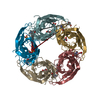
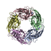
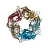
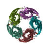
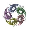
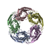
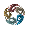
| Title | Crystal Structure of AChBP from Bulinus truncatus revals the conserved structural scaffold and sites of variation in nicotinic acetylcholine receptors | ||||||
 Components Components | ACETYLCHOLINE-BINDING PROTEIN | ||||||
 Keywords Keywords | GLYCOPROTEIN / GLYCPROTEIN / IGG FOLD / IMMUNOGLOBULIN DOMAIN / PENTAMER | ||||||
| Function / homology | Acetylcholine Binding Protein; Chain: A, / Neurotransmitter-gated ion-channel ligand-binding domain / Distorted Sandwich / Mainly Beta Function and homology information Function and homology information | ||||||
| Biological species |  BULINUS TRUNCATUS (invertebrata) BULINUS TRUNCATUS (invertebrata) | ||||||
| Method |  X-RAY DIFFRACTION / SYNCHROTRON / MOLECULAR REPLACEMENT / Resolution: 2 Å X-RAY DIFFRACTION / SYNCHROTRON / MOLECULAR REPLACEMENT / Resolution: 2 Å | ||||||
 Authors Authors | Celie, P.H.N. / Klaassen, R.V. / Van Rossum-Fikkert, S.E. / Van Elk, R. / Van Nierop, P. / Smit, A.B. / Sixma, T.K. | ||||||
 Citation Citation | Journal: J.Biol.Chem. / Year: 2005 Title: Crystal Structure of Acetylcholine-Binding Protein from Bulinus Truncatus Reveals the Conserved Structural Scaffold and Sites of Variation in Nicotinic Acetylcholine Receptors. Authors: Celie, P.H.N. / Klaassen, R.V. / Van Rossum-Fikkert, S.E. / Van Elk, R. / Van Nierop, P. / Smit, A.B. / Sixma, T.K. | ||||||
| History |
| ||||||
| Remark 700 | SHEET THE SHEET STRUCTURE OF THIS MOLECULE IS BIFURCATED. IN ORDER TO REPRESENT THIS FEATURE IN ... SHEET THE SHEET STRUCTURE OF THIS MOLECULE IS BIFURCATED. IN ORDER TO REPRESENT THIS FEATURE IN THE SHEET RECORDS BELOW, TWO SHEETS ARE DEFINED. |
- Structure visualization
| Structure viewer | Molecule: MolmilJmol/JSmol |
|---|
- Downloads & links
-Download
| PDBx/mmCIF format | 2bj0.cif.gz | 214.6 KB | Display | PDBx/mmCIF format |
|---|---|---|---|---|
| PDB format | pdb2bj0.ent.gz | 175 KB | Display | PDB format |
| PDBx/mmJSON format | 2bj0.json.gz | Tree view | PDBx/mmJSON format | |
| Others |  Other downloads Other downloads |
-Validation report
| Arichive directory | https://data.pdbj.org/pub/pdb/validation_reports/bj/2bj0ftp://data.pdbj.org/pub/pdb/validation_reports/bj/2bj0 | HTTPS FTP |
|---|
-Related structure data
| Related structure data | 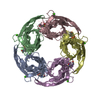 1i9bS S: Starting model for refinement |
|---|---|
| Similar structure data |


-Links
 PDBj
PDBj- Assembly
| Deposited unit | 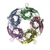
| ||||||||||||||||||||
|---|---|---|---|---|---|---|---|---|---|---|---|---|---|---|---|---|---|---|---|---|---|
| 1 |
| ||||||||||||||||||||
| Unit cell |
| ||||||||||||||||||||
| Components on special symmetry positions |
| ||||||||||||||||||||
| Noncrystallographic symmetry (NCS) | NCS oper:
|
-Components
| #1: Protein | Mass: 22908.893 Da / Num. of mol.: 5 Source method: isolated from a genetically manipulated source Details: CAPS BUFFER MOLECULE IS COCRYSTALLIZED IN THE LIGAND BINDING-SITE. CAPS COULD BE BUILT IN 4 OUT OF 5 BINDING SITES WITHIN THE PENTAMER Source: (gene. exp.) BULINUS TRUNCATUS (invertebrata) / Cell: GLIAL / Plasmid: PFASTBACI / Cell line (production host): SF9 / Production host:   SPODOPTERA FRUGIPERDA (fall armyworm) SPODOPTERA FRUGIPERDA (fall armyworm)#2: Chemical | ChemComp-CXS /   Mass: 221.317 Da / Num. of mol.: 4 / Source method: obtained synthetically / Formula: C9H19NO3S / Comment: pH buffer*YM Mass: 221.317 Da / Num. of mol.: 4 / Source method: obtained synthetically / Formula: C9H19NO3S / Comment: pH buffer*YM#3: Water | ChemComp-HOH / |  Mass: 18.015 Da / Num. of mol.: 424 / Source method: isolated from a natural source / Formula: H2O Mass: 18.015 Da / Num. of mol.: 424 / Source method: isolated from a natural source / Formula: H2OHas protein modification | Y | Sequence details | ACHBP FROM BULINUS TRUNCATUS CONTAINS SIGNAL SEQUENCE (MAELRGIILL LCTIAFHVSH G) THAT PRECEDES ...ACHBP FROM BULINUS TRUNCATUS CONTAINS SIGNAL SEQUENCE (MAELRGIILL | |
|---|
-Experimental details
-Experiment
| Experiment | Method: X-RAY DIFFRACTION / Number of used crystals: 1 |
|---|
- Sample preparation
| Crystal | Density Matthews: 2.9 Å3/Da / Density % sol: 57.7 % |
|---|---|
| Crystal grow | pH: 10.5 Details: 0.1 M CAPS PH10.5, 0.2 M LITHIUMSULFATE, 2 M AMMONIUM SULFATE, pH 10.50 |
-Data collection
| Diffraction | Mean temperature: 100 K |
|---|---|
| Diffraction source | Source: SYNCHROTRON / Site: ESRF  / Beamline: ID14-4 / Wavelength: 0.9393 / Beamline: ID14-4 / Wavelength: 0.9393 |
| Detector | Type: ADSC CCD / Detector: CCD / Date: Mar 19, 2003 |
| Radiation | Protocol: SINGLE WAVELENGTH / Monochromatic (M) / Laue (L): M / Scattering type: x-ray |
| Radiation wavelength | Wavelength: 0.9393 Å / Relative weight: 1 |
| Reflection | Resolution: 2→44.72 Å / Num. obs: 93560 / % possible obs: 98 % / Observed criterion σ(I): 1.7 / Redundancy: 5.1 % / Rmerge(I) obs: 0.13 / Net I/σ(I): 12 |
| Reflection shell | Resolution: 2→2.07 Å / Redundancy: 3.4 % / Rmerge(I) obs: 0.77 / Mean I/σ(I) obs: 1.7 / % possible all: 89 |
- Processing
| Software |
| ||||||||||||||||||||||||||||||||||||||||||||||||||||||||||||||||||||||||||||||||||||||||||||||||||||||||||||||||||||||||||||||||||||||||||||||||||||||||||||||||||||||||||||||||||||||
|---|---|---|---|---|---|---|---|---|---|---|---|---|---|---|---|---|---|---|---|---|---|---|---|---|---|---|---|---|---|---|---|---|---|---|---|---|---|---|---|---|---|---|---|---|---|---|---|---|---|---|---|---|---|---|---|---|---|---|---|---|---|---|---|---|---|---|---|---|---|---|---|---|---|---|---|---|---|---|---|---|---|---|---|---|---|---|---|---|---|---|---|---|---|---|---|---|---|---|---|---|---|---|---|---|---|---|---|---|---|---|---|---|---|---|---|---|---|---|---|---|---|---|---|---|---|---|---|---|---|---|---|---|---|---|---|---|---|---|---|---|---|---|---|---|---|---|---|---|---|---|---|---|---|---|---|---|---|---|---|---|---|---|---|---|---|---|---|---|---|---|---|---|---|---|---|---|---|---|---|---|---|---|---|
| Refinement | Method to determine structure: MOLECULAR REPLACEMENT Starting model: PDB ENTRY 1I9B Resolution: 2→12 Å / Cor.coef. Fo:Fc: 0.951 / Cor.coef. Fo:Fc free: 0.93 / SU B: 4.748 / SU ML: 0.13 / Cross valid method: THROUGHOUT / ESU R: 0.171 / ESU R Free: 0.159 / Stereochemistry target values: MAXIMUM LIKELIHOOD / Details: HYDROGENS HAVE BEEN ADDED IN THE RIDING POSITIONS.
| ||||||||||||||||||||||||||||||||||||||||||||||||||||||||||||||||||||||||||||||||||||||||||||||||||||||||||||||||||||||||||||||||||||||||||||||||||||||||||||||||||||||||||||||||||||||
| Solvent computation | Ion probe radii: 0.8 Å / Shrinkage radii: 0.8 Å / VDW probe radii: 1.2 Å / Solvent model: BABINET MODEL WITH MASK | ||||||||||||||||||||||||||||||||||||||||||||||||||||||||||||||||||||||||||||||||||||||||||||||||||||||||||||||||||||||||||||||||||||||||||||||||||||||||||||||||||||||||||||||||||||||
| Displacement parameters | Biso mean: 34.23 Å2
| ||||||||||||||||||||||||||||||||||||||||||||||||||||||||||||||||||||||||||||||||||||||||||||||||||||||||||||||||||||||||||||||||||||||||||||||||||||||||||||||||||||||||||||||||||||||
| Refinement step | Cycle: LAST / Resolution: 2→12 Å
| ||||||||||||||||||||||||||||||||||||||||||||||||||||||||||||||||||||||||||||||||||||||||||||||||||||||||||||||||||||||||||||||||||||||||||||||||||||||||||||||||||||||||||||||||||||||
| Refine LS restraints |
|