[English] 日本語
Yorodumi
- PDB-2bhu: Crystal structure of Deinococcus radiodurans maltooligosyltrehalo... -

+
Open data


ID or keywords:

Loading...

-
Basic information

Entry
Database: PDB / ID: 2bhu
TitleCrystal structure of Deinococcus radiodurans maltooligosyltrehalose trehalohydrolase
ComponentsMALTOOLIGOSYLTREHALOSE TREHALOHYDROLASE
KeywordsHYDROLASE / TREHALOSE / ALPHA-AMYLASE / PROTEIN-CARBOHYDRATE COMPLEX / DESICCATION RESISTANCE
Function / homology
Function and homology information


4-alpha-D-{(1->4)-alpha-D-glucano}trehalose trehalohydrolase / 4-alpha-D-(1->4)-alpha-D-glucanotrehalose trehalohydrolase activity / trehalose biosynthetic process / cytoplasm
Similarity search - Function
Malto-oligosyltrehalose trehalohydrolase, E-set domain superfamily / Malto-oligosyltrehalose trehalohydrolase / Glycosyl hydrolase, C-terminal (DUF3459) / Domain of unknown function (DUF3459) / Glycoside hydrolase, family 13, N-terminal / Carbohydrate-binding module 48 (Isoamylase N-terminal domain) / Alpha amylase, catalytic domain / Glycosyl hydrolase, family 13, catalytic domain / Alpha-amylase domain / Golgi alpha-mannosidase II ...Malto-oligosyltrehalose trehalohydrolase, E-set domain superfamily / Malto-oligosyltrehalose trehalohydrolase / Glycosyl hydrolase, C-terminal (DUF3459) / Domain of unknown function (DUF3459) / Glycoside hydrolase, family 13, N-terminal / Carbohydrate-binding module 48 (Isoamylase N-terminal domain) / Alpha amylase, catalytic domain / Glycosyl hydrolase, family 13, catalytic domain / Alpha-amylase domain / Golgi alpha-mannosidase II / Glycosyl hydrolase, all-beta / Glycosidases / Immunoglobulin E-set / Glycoside hydrolase superfamily / Immunoglobulins / TIM Barrel / Alpha-Beta Barrel / Immunoglobulin-like fold / Immunoglobulin-like / Sandwich / Mainly Beta / Alpha Beta
Similarity search - Domain/homology
TRIETHYLENE GLYCOL / Malto-oligosyltrehalose trehalohydrolase
Similarity search - Component
Biological speciesDEINOCOCCUS RADIODURANS (radioresistant)
MethodX-RAY DIFFRACTION / SYNCHROTRON / SIRAS / Resolution: 1.1 Å
AuthorsTimmins, J. / Leiros, H.-K.S. / Leonard, G. / Leiros, I. / McSweeney, S.
CitationJournal: J.Mol.Biol. / Year: 2005
Title: Crystal Structure of Maltooligosyltrehalose Trehalohydrolase from Deinococcus Radiodurans in Complex with Disaccharides
Authors: Timmins, J. / Leiros, H.-K.S. / Leonard, G. / Leiros, I. / Mcsweeney, S.
History
DepositionJan 18, 2005Deposition site: PDBE / Processing site: PDBE
Revision 1.0Mar 31, 2005Provider: repository / Type: Initial release
Revision 1.1May 8, 2011Group: Version format compliance
Revision 1.2Jul 13, 2011Group: Version format compliance
Remark 700 
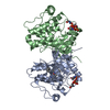
SHEET DETERMINATION METHOD: DSSP THE SHEETS PRESENTED AS "AD" IN EACH CHAIN ON SHEET RECORDS BELOW ... SHEET DETERMINATION METHOD: DSSP THE SHEETS PRESENTED AS "AD" IN EACH CHAIN ON SHEET RECORDS BELOW IS ACTUALLY AN 8-STRANDED BARREL THIS IS REPRESENTED BY A 9-STRANDED SHEET IN WHICH THE FIRST AND LAST STRANDS ARE IDENTICAL.

-
Structure visualization

Structure viewerMolecule:
MolmilJmol/JSmol

Downloads & links

-
Assembly

Deposited unit
A: MALTOOLIGOSYLTREHALOSE TREHALOHYDROLASE
hetero molecules


Theoretical massNumber of molelcules
Total (without water)67,4984
Polymers67,2021
Non-polymers2973
Water20,3571130
1


  • Idetical with deposited unit
  • defined by author&software
TypeNameSymmetry operationNumber
identity operation1_555x,y,z1
MethodPQS
Unit cell
Length a, b, c (Å)59.580, 66.620, 152.510
Angle α, β, γ (deg.)90.00, 90.00, 90.00
Int Tables number19
Space group name H-MP212121

-
Components

#1: Protein MALTOOLIGOSYLTREHALOSE TREHALOHYDROLASE


Mass: 67201.711 Da / Num. of mol.: 1 / Mutation: YES
Source method: isolated from a genetically manipulated source
Source: (gene. exp.) DEINOCOCCUS RADIODURANS (radioresistant)
Strain: R1 / Plasmid: PDEST17 / Production host: ESCHERICHIA COLI (E. coli) / Strain (production host): BL21(DE3) / References: UniProt: Q9RX51, alpha-amylase
#2: Chemical ChemComp-TRS / 2-AMINO-2-HYDROXYMETHYL-PROPANE-1,3-DIOL / TRIS BUFFER / Tris


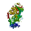
Mass: 122.143 Da / Num. of mol.: 1 / Source method: obtained synthetically / Formula: C4H12NO3 / Comment: pH buffer*YM
#3: Chemical ChemComp-PGE / TRIETHYLENE GLYCOL / Polyethylene glycol


Mass: 150.173 Da / Num. of mol.: 1 / Source method: obtained synthetically / Formula: C6H14O4
#4: Chemical ChemComp-MG / MAGNESIUM ION


Mass: 24.305 Da / Num. of mol.: 1 / Source method: obtained synthetically / Formula: Mg
#5: Water ChemComp-HOH / water / Water


Mass: 18.015 Da / Num. of mol.: 1130 / Source method: isolated from a natural source / Formula: H2O
Compound detailsCHAIN A INSERTION MUTATION BETWEEN RESIDUES 430 AND 431 IN THE UNIPROT ENTRY. RESIDUES THR431 AND ...CHAIN A INSERTION MUTATION BETWEEN RESIDUES 430 AND 431 IN THE UNIPROT ENTRY. RESIDUES THR431 AND LEU432 INSERTED AT THIS POSITION.

-
Experimental details

-
Experiment

ExperimentMethod: X-RAY DIFFRACTION / Number of used crystals: 1

-
Sample preparation

CrystalDensity Matthews: 1.75 Å3/Da / Density % sol: 29.22 %
Crystal growpH: 8.5
Details: 20% PEG 2000 MME, 0.1 M TRIS-HCL PH8.5, 0.1 M MGCL2, pH 8.50

-
Data collection

DiffractionMean temperature: 100 K
Diffraction sourceSource: SYNCHROTRON / Site: ESRF / Beamline: ID29 / Wavelength: 1.005
DetectorDate: Jul 30, 2003
RadiationProtocol: SINGLE WAVELENGTH / Monochromatic (M) / Laue (L): M / Scattering type: x-ray
Radiation wavelengthWavelength: 1.005 Å / Relative weight: 1
ReflectionResolution: 1.1→46.93 Å / Num. obs: 216600 / % possible obs: 88.3 % / Observed criterion σ(I): 0 / Redundancy: 2.6 % / Rmerge(I) obs: 0.08 / Net I/σ(I): 7.1
Reflection shellResolution: 1.1→1.16 Å / Redundancy: 1.8 % / Rmerge(I) obs: 0.39 / Mean I/σ(I) obs: 2.3 / % possible all: 82.9

-
Processing

Software
NameVersionClassification
REFMAC5.2.0003refinement
XDSdata reduction
XDSdata scaling
SOLVEphasing
RefinementMethod to determine structure: SIRAS / Resolution: 1.1→44.41 Å / Cor.coef. Fo:Fc: 0.981 / Cor.coef. Fo:Fc free: 0.974 / SU B: 0.99 / SU ML: 0.021 / Cross valid method: THROUGHOUT / ESU R: 0.03 / ESU R Free: 0.032 / Stereochemistry target values: MAXIMUM LIKELIHOOD
Details: HYDROGENS HAVE BEEN ADDED IN THE RIDING POSITIONS. WARNING: THIS ENTRY CONTAINS ATOMS THAT HAVE BEEN REFINED WITH AN OCCUPANCY OF 0.00.
RfactorNum. reflection% reflectionSelection details
Rfree0.159 10891 5 %RANDOM
Rwork0.128 ---
obs0.129 205610 88.2 %-
Solvent computationIon probe radii: 0.8 Å / Shrinkage radii: 0.8 Å / VDW probe radii: 1.2 Å / Solvent model: MASK
Displacement parametersBiso mean: 9.11 Å2
Baniso -1Baniso -2Baniso -3
1-0.39 Å20 Å20 Å2
2---0.39 Å20 Å2
3---0 Å2
Refinement stepCycle: LAST / Resolution: 1.1→44.41 Å
ProteinNucleic acidLigandSolventTotal
Num. atoms4601 0 19 1130 5750
Refine LS restraints
Refine-IDTypeDev idealDev ideal targetNumber
X-RAY DIFFRACTIONr_bond_refined_d0.0170.0215187
X-RAY DIFFRACTIONr_bond_other_d0.0020.024551
X-RAY DIFFRACTIONr_angle_refined_deg1.7971.9297122
X-RAY DIFFRACTIONr_angle_other_deg0.951310547
X-RAY DIFFRACTIONr_dihedral_angle_1_deg5.9745672
X-RAY DIFFRACTIONr_dihedral_angle_2_deg35.7922.984258
X-RAY DIFFRACTIONr_dihedral_angle_3_deg11.58815770
X-RAY DIFFRACTIONr_dihedral_angle_4_deg17.391547
X-RAY DIFFRACTIONr_chiral_restr0.1260.2735
X-RAY DIFFRACTIONr_gen_planes_refined0.0110.026135
X-RAY DIFFRACTIONr_gen_planes_other0.0030.021171
X-RAY DIFFRACTIONr_nbd_refined0.2650.21126
X-RAY DIFFRACTIONr_nbd_other0.2060.25146
X-RAY DIFFRACTIONr_nbtor_refined0.1860.22582
X-RAY DIFFRACTIONr_nbtor_other0.0870.23006
X-RAY DIFFRACTIONr_xyhbond_nbd_refined0.1960.2842
X-RAY DIFFRACTIONr_xyhbond_nbd_other
X-RAY DIFFRACTIONr_metal_ion_refined
X-RAY DIFFRACTIONr_metal_ion_other
X-RAY DIFFRACTIONr_symmetry_vdw_refined1.0750.241
X-RAY DIFFRACTIONr_symmetry_vdw_other0.5670.2175
X-RAY DIFFRACTIONr_symmetry_hbond_refined0.2120.2104
X-RAY DIFFRACTIONr_symmetry_hbond_other
X-RAY DIFFRACTIONr_symmetry_metal_ion_refined
X-RAY DIFFRACTIONr_symmetry_metal_ion_other
X-RAY DIFFRACTIONr_mcbond_it1.9411.54039
X-RAY DIFFRACTIONr_mcbond_other
X-RAY DIFFRACTIONr_mcangle_it2.35325127
X-RAY DIFFRACTIONr_mcangle_other
X-RAY DIFFRACTIONr_scbond_it3.11632357
X-RAY DIFFRACTIONr_scbond_other
X-RAY DIFFRACTIONr_scangle_it3.9334.51995
X-RAY DIFFRACTIONr_scangle_other
X-RAY DIFFRACTIONr_long_range_B_refined
X-RAY DIFFRACTIONr_long_range_B_other
X-RAY DIFFRACTIONr_rigid_bond_restr
X-RAY DIFFRACTIONr_sphericity_free
X-RAY DIFFRACTIONr_sphericity_bonded
LS refinement shellResolution: 1.1→1.13 Å / Total num. of bins used: 20 /
RfactorNum. reflection
Rfree0.279 707
Rwork0.258 13628

+
About Yorodumi

-
News

-
Feb 9, 2022. New format data for meta-information of EMDB entries

New format data for meta-information of EMDB entries

  • Version 3 of the EMDB header file is now the official format.
  • The previous official version 1.9 will be removed from the archive.

Related info.:EMDB header

External links:wwPDB to switch to version 3 of the EMDB data model

-
Aug 12, 2020. Covid-19 info

Covid-19 info

URL: https://pdbj.org/emnavi/covid19.php

New page: Covid-19 featured information page in EM Navigator.

Related info.:Covid-19 info / Mar 5, 2020. Novel coronavirus structure data

+
Mar 5, 2020. Novel coronavirus structure data

Novel coronavirus structure data

Related info.:Yorodumi Speices / Aug 12, 2020. Covid-19 info

External links:COVID-19 featured content - PDBj / Molecule of the Month (242):Coronavirus Proteases

+
Jan 31, 2019. EMDB accession codes are about to change! (news from PDBe EMDB page)

EMDB accession codes are about to change! (news from PDBe EMDB page)

  • The allocation of 4 digits for EMDB accession codes will soon come to an end. Whilst these codes will remain in use, new EMDB accession codes will include an additional digit and will expand incrementally as the available range of codes is exhausted. The current 4-digit format prefixed with “EMD-” (i.e. EMD-XXXX) will advance to a 5-digit format (i.e. EMD-XXXXX), and so on. It is currently estimated that the 4-digit codes will be depleted around Spring 2019, at which point the 5-digit format will come into force.
  • The EM Navigator/Yorodumi systems omit the EMD- prefix.

Related info.:Q: What is EMD? / ID/Accession-code notation in Yorodumi/EM Navigator

External links:EMDB Accession Codes are Changing Soon! / Contact to PDBj

+
Jul 12, 2017. Major update of PDB

Major update of PDB

  • wwPDB released updated PDB data conforming to the new PDBx/mmCIF dictionary.
  • This is a major update changing the version number from 4 to 5, and with Remediation, in which all the entries are updated.
  • In this update, many items about electron microscopy experimental information are reorganized (e.g. em_software).
  • Now, EM Navigator and Yorodumi are based on the updated data.

External links:wwPDB Remediation / Enriched Model Files Conforming to OneDep Data Standards Now Available in the PDB FTP Archive

-
Yorodumi

Thousand views of thousand structures

  • Yorodumi is a browser for structure data from EMDB, PDB, SASBDB, etc.
  • This page is also the successor to EM Navigator detail page, and also detail information page/front-end page for Omokage search.
  • The word "yorodu" (or yorozu) is an old Japanese word meaning "ten thousand". "mi" (miru) is to see.

Related info.:EMDB / PDB / SASBDB / Comparison of 3 databanks / Yorodumi Search / Aug 31, 2016. New EM Navigator & Yorodumi / Yorodumi Papers / Jmol/JSmol / Function and homology information / Changes in new EM Navigator and Yorodumi

Read more