 Movie
Movie Controller

[English] 日本語
 Yorodumi
- PDB-1c8u: CRYSTAL STRUCTURE OF THE E.COLI THIOESTERASE II, A HOMOLOGUE OF T... -
+ Open data

- Basic information
| Entry | Database: PDB / ID: 1c8u | ||||||
|---|---|---|---|---|---|---|---|
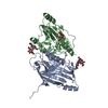
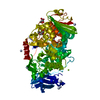
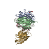
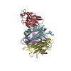
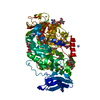
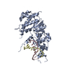
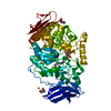
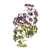
| Title | CRYSTAL STRUCTURE OF THE E.COLI THIOESTERASE II, A HOMOLOGUE OF THE HUMAN NEF-BINDING ENZYME | ||||||
 Components Components | ACYL-COA THIOESTERASE II | ||||||
 Keywords Keywords | HYDROLASE / INTERNAL REPEATS | ||||||
| Function / homology |  Function and homology information Function and homology informationacyl-CoA hydrolase / acyl-CoA metabolic process / fatty acyl-CoA hydrolase activity / fatty acid catabolic process / Hydrolases; Acting on ester bonds; Thioester hydrolases / protein homodimerization activity / identical protein binding / cytosol Similarity search - Function | ||||||
| Biological species |  | ||||||
| Method |  X-RAY DIFFRACTION / SYNCHROTRON / Resolution: 1.9 Å X-RAY DIFFRACTION / SYNCHROTRON / Resolution: 1.9 Å | ||||||
 Authors Authors | Li, J. / Derewenda, Z.S. | ||||||
 Citation Citation | Journal: Nat.Struct.Biol. / Year: 2000 Title: Crystal structure of the Escherichia coli thioesterase II, a homolog of the human Nef binding enzyme. Authors: Li, J. / Derewenda, U. / Dauter, Z. / Smith, S. / Derewenda, Z.S. #1: Journal: J.Mol.Biol. / Year: 1994Title: Crystallization of Thioesterase II from Escherichia Coli Authors: Swenson, L. / Green, R. / Smith, S. / Derewenda, Z.S. | ||||||
| History |
|
- Structure visualization
| Structure viewer | Molecule: MolmilJmol/JSmol |
|---|
- Downloads & links
-Download
| PDBx/mmCIF format | 1c8u.cif.gz | 134.8 KB | Display | PDBx/mmCIF format |
|---|---|---|---|---|
| PDB format | pdb1c8u.ent.gz | 105.5 KB | Display | PDB format |
| PDBx/mmJSON format | 1c8u.json.gz | Tree view | PDBx/mmJSON format | |
| Others |  Other downloads Other downloads |
-Validation report
| Arichive directory | https://data.pdbj.org/pub/pdb/validation_reports/c8/1c8uftp://data.pdbj.org/pub/pdb/validation_reports/c8/1c8u | HTTPS FTP |
|---|
-Related structure data
| Similar structure data |
|---|


-Links
 PDBj
PDBj
- Assembly
| Deposited unit | 
| ||||||||||
|---|---|---|---|---|---|---|---|---|---|---|---|
| 1 |
| ||||||||||
| Unit cell |
|
-Components
| #1: Protein | Mass: 31873.062 Da / Num. of mol.: 2 Source method: isolated from a genetically manipulated source Source: (gene. exp.) References: UniProt: P23911, UniProt: P0AGG2*PLUS, Hydrolases; Acting on ester bonds; Thioester hydrolases #2: Chemical |   Mass: 229.402 Da / Num. of mol.: 2 / Source method: obtained synthetically / Formula: C14H31NO / Comment: LDAO, detergent*YM Mass: 229.402 Da / Num. of mol.: 2 / Source method: obtained synthetically / Formula: C14H31NO / Comment: LDAO, detergent*YM#3: Water | ChemComp-HOH / |  Mass: 18.015 Da / Num. of mol.: 533 / Source method: isolated from a natural source / Formula: H2O Mass: 18.015 Da / Num. of mol.: 533 / Source method: isolated from a natural source / Formula: H2O |
|---|
-Experimental details
-Experiment
| Experiment | Method: X-RAY DIFFRACTION / Number of used crystals: 1 |
|---|
- Sample preparation
| Crystal | Density Matthews: 3.73 Å3/Da / Density % sol: 66.99 % | ||||||||||||||||||||||||||||||||||||
|---|---|---|---|---|---|---|---|---|---|---|---|---|---|---|---|---|---|---|---|---|---|---|---|---|---|---|---|---|---|---|---|---|---|---|---|---|---|
| Crystal grow | Temperature: 293 K / Method: vapor diffusion, sitting drop / pH: 6.5 Details: NACL, NAOAC, LDAO, pH 6.5, VAPOR DIFFUSION, SITTING DROP, temperature 293K | ||||||||||||||||||||||||||||||||||||
| Crystal grow | *PLUS pH: 7.5 | ||||||||||||||||||||||||||||||||||||
| Components of the solutions | *PLUS
|
-Data collection
| Diffraction | Mean temperature: 120 K |
|---|---|
| Diffraction source | Source: SYNCHROTRON / Site: EMBL/DESY, HAMBURG  / Beamline: X11 / Wavelength: 0.9096 / Beamline: X11 / Wavelength: 0.9096 |
| Detector | Type: MARRESEARCH / Detector: IMAGE PLATE / Date: Nov 10, 1997 |
| Radiation | Protocol: SINGLE WAVELENGTH / Monochromatic (M) / Laue (L): M / Scattering type: x-ray |
| Radiation wavelength | Wavelength: 0.9096 Å / Relative weight: 1 |
| Reflection | Resolution: 1.9→20 Å / Num. all: 74901 / Num. obs: 74732 / % possible obs: 99.8 % / Observed criterion σ(I): 1 / Redundancy: 4.7 % / Biso Wilson estimate: 24 Å2 / Rmerge(I) obs: 0.059 / Net I/σ(I): 10.8 |
| Reflection shell | Resolution: 1.9→1.97 Å / Redundancy: 4.1 % / Rmerge(I) obs: 0.425 / % possible all: 99.4 |
| Reflection | *PLUS Num. obs: 74901 / % possible obs: 99.7 % / Num. measured all: 353158 |
| Reflection shell | *PLUS % possible obs: 99.4 % |
- Processing
| Software |
| ||||||||||||||||||||||||||||||||||||||||||||||||||||||||||||
|---|---|---|---|---|---|---|---|---|---|---|---|---|---|---|---|---|---|---|---|---|---|---|---|---|---|---|---|---|---|---|---|---|---|---|---|---|---|---|---|---|---|---|---|---|---|---|---|---|---|---|---|---|---|---|---|---|---|---|---|---|---|
| Refinement | Resolution: 1.9→20 Å / Cross valid method: THROUGHOUT / σ(F): 0 / σ(I): 0 / Stereochemistry target values: ENGH & HUBER
| ||||||||||||||||||||||||||||||||||||||||||||||||||||||||||||
| Refinement step | Cycle: LAST / Resolution: 1.9→20 Å
| ||||||||||||||||||||||||||||||||||||||||||||||||||||||||||||
| Refine LS restraints |
| ||||||||||||||||||||||||||||||||||||||||||||||||||||||||||||
| Software | *PLUS Name: 'CNS' / Classification: refinement | ||||||||||||||||||||||||||||||||||||||||||||||||||||||||||||
| Refinement | *PLUS Rfactor Rfree: 0.248 / Rfactor Rwork: 0.218 | ||||||||||||||||||||||||||||||||||||||||||||||||||||||||||||
| Solvent computation | *PLUS | ||||||||||||||||||||||||||||||||||||||||||||||||||||||||||||
| Displacement parameters | *PLUS |
