 Movie
Movie Controller

[English] 日本語
 Yorodumi
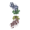
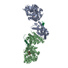
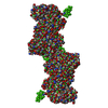
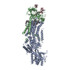
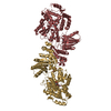
- PDB-2bdn: Crystal structure of human MCP-1 bound to a blocking antibody, 11K2 -
+ Open data

- Basic information
| Entry | Database: PDB / ID: 2bdn | ||||||
|---|---|---|---|---|---|---|---|
| Title | Crystal structure of human MCP-1 bound to a blocking antibody, 11K2 | ||||||
 Components Components |
| ||||||
 Keywords Keywords | IMMUNE SYSTEM / antibody-antigen complex | ||||||
| Function / homology |  Function and homology information Function and homology informationhelper T cell extravasation / chemokine (C-C motif) ligand 2 signaling pathway / CCR2 chemokine receptor binding / negative regulation of natural killer cell chemotaxis / astrocyte cell migration / chemokine receptor binding / ATF4 activates genes in response to endoplasmic reticulum stress / CCR chemokine receptor binding / negative regulation of glial cell apoptotic process / positive regulation of apoptotic cell clearance ...helper T cell extravasation / chemokine (C-C motif) ligand 2 signaling pathway / CCR2 chemokine receptor binding / negative regulation of natural killer cell chemotaxis / astrocyte cell migration / chemokine receptor binding / ATF4 activates genes in response to endoplasmic reticulum stress / CCR chemokine receptor binding / negative regulation of glial cell apoptotic process / positive regulation of apoptotic cell clearance / NFE2L2 regulating inflammation associated genes / cellular homeostasis / eosinophil chemotaxis / positive regulation of glutamate receptor signaling pathway / negative regulation of vascular endothelial cell proliferation / chemokine activity / Chemokine receptors bind chemokines / negative regulation of G1/S transition of mitotic cell cycle / positive regulation of macrophage chemotaxis / chemoattractant activity / positive regulation of calcium ion import / Interleukin-10 signaling / humoral immune response / macrophage chemotaxis / monocyte chemotaxis / positive regulation of endothelial cell apoptotic process / cellular response to interleukin-1 / G protein-coupled receptor signaling pathway, coupled to cyclic nucleotide second messenger / cell surface receptor signaling pathway via JAK-STAT / positive regulation of synaptic transmission, glutamatergic / cytoskeleton organization / cellular response to fibroblast growth factor stimulus / response to bacterium / sensory perception of pain / viral genome replication / animal organ morphogenesis / chemokine-mediated signaling pathway / cellular response to type II interferon / cellular response to tumor necrosis factor / chemotaxis / positive regulation of T cell activation / cytokine-mediated signaling pathway / regulation of cell shape / antimicrobial humoral immune response mediated by antimicrobial peptide / positive regulation of cytosolic calcium ion concentration / cellular response to lipopolysaccharide / angiogenesis / Interleukin-4 and Interleukin-13 signaling / negative regulation of neuron apoptotic process / protein phosphorylation / protein kinase activity / cell surface receptor signaling pathway / cell adhesion / positive regulation of cell migration / inflammatory response / G protein-coupled receptor signaling pathway / signaling receptor binding / positive regulation of gene expression / signal transduction / : / extracellular region / membrane Similarity search - Function | ||||||
| Biological species |  | ||||||
| Method |  X-RAY DIFFRACTION / SYNCHROTRON / MOLECULAR REPLACEMENT / Resolution: 2.53 Å X-RAY DIFFRACTION / SYNCHROTRON / MOLECULAR REPLACEMENT / Resolution: 2.53 Å | ||||||
 Authors Authors | Boriack-Sjodin, P.A. / Rushe, M. / Reid, C. / Jarpe, M. / van Vlijmen, H. / Bailly, V. | ||||||
 Citation Citation | Journal: Protein Eng.Des.Sel. / Year: 2006 Title: Structure activity relationships of monocyte chemoattractant proteins in complex with a blocking antibody. Authors: Reid, C. / Rushe, M. / Jarpe, M. / van Vlijmen, H. / Dolinski, B. / Qian, F. / Cachero, T.G. / Cuervo, H. / Yanachkova, M. / Nwankwo, C. / Wang, X. / Etienne, N. / Garber, E. / Bailly, V. / ...Authors: Reid, C. / Rushe, M. / Jarpe, M. / van Vlijmen, H. / Dolinski, B. / Qian, F. / Cachero, T.G. / Cuervo, H. / Yanachkova, M. / Nwankwo, C. / Wang, X. / Etienne, N. / Garber, E. / Bailly, V. / de Fougerolles, A. / Boriack-Sjodin, P.A. | ||||||
| History |
| ||||||
| Remark 999 | Sequence No suitable sequence database reference was available for the proteins in chains L and H ...Sequence No suitable sequence database reference was available for the proteins in chains L and H at the time of processing this file. |
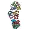
- Structure visualization
| Structure viewer | Molecule: MolmilJmol/JSmol |
|---|
- Downloads & links
-Download
| PDBx/mmCIF format | 2bdn.cif.gz | 107.7 KB | Display | PDBx/mmCIF format |
|---|---|---|---|---|
| PDB format | pdb2bdn.ent.gz | 83.3 KB | Display | PDB format |
| PDBx/mmJSON format | 2bdn.json.gz | Tree view | PDBx/mmJSON format | |
| Others |  Other downloads Other downloads |
-Validation report
| Arichive directory | https://data.pdbj.org/pub/pdb/validation_reports/bd/2bdnftp://data.pdbj.org/pub/pdb/validation_reports/bd/2bdn | HTTPS FTP |
|---|
-Related structure data
| Related structure data |  11k2S S: Starting model for refinement |
|---|---|
| Similar structure data |


-Links
 PDBj
PDBj


- Assembly
| Deposited unit | 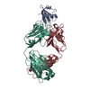
| ||||||||
|---|---|---|---|---|---|---|---|---|---|
| 1 | 
| ||||||||
| Unit cell |
| ||||||||
| Components on special symmetry positions |
| ||||||||
| Details | The MCP-1 dimer is generated by the symmetry operator x, -y, -z and translation of the resulting symmetry protein by 0 0 1 fractional (ie, translation along the Z axis by 1 unit cell length). |
-Components
| #1: Protein | Mass: 8699.045 Da / Num. of mol.: 1 / Fragment: monocyte chemotactic protein -1 / Source method: obtained synthetically Details: Chemically synthesized. This sequence occurs naturally in humans References: UniProt: P13500 |
|---|---|
| #2: Antibody | Mass: 23574.939 Da / Num. of mol.: 1 / Fragment: fab fragment Source method: isolated from a genetically manipulated source Source: (gene. exp.) |
| #3: Antibody | Mass: 23122.873 Da / Num. of mol.: 1 / Fragment: fab fragment Source method: isolated from a genetically manipulated source Source: (gene. exp.) |
| #4: Water | ChemComp-HOH /  Mass: 18.015 Da / Num. of mol.: 43 / Source method: isolated from a natural source / Formula: H2O Mass: 18.015 Da / Num. of mol.: 43 / Source method: isolated from a natural source / Formula: H2O |
| Has protein modification | Y |
-Experimental details
-Experiment
| Experiment | Method: X-RAY DIFFRACTION / Number of used crystals: 1 |
|---|
- Sample preparation
| Crystal | Density Matthews: 3.11 Å3/Da / Density % sol: 60.44 % |
|---|---|
| Crystal grow | Temperature: 298 K / Method: vapor diffusion, hanging drop / pH: 7.5 Details: 13% PEG 4000, 100 mM Hepes, pH 7.5, 30 mM glycl-glycl-glycine, VAPOR DIFFUSION, HANGING DROP, temperature 298K |
-Data collection
| Diffraction | Mean temperature: 100 K |
|---|---|
| Diffraction source | Source: SYNCHROTRON / Site: NSLS  / Beamline: X4A / Wavelength: 0.97945 Å / Beamline: X4A / Wavelength: 0.97945 Å |
| Detector | Type: ADSC QUANTUM 4 / Detector: CCD / Date: Nov 19, 2002 |
| Radiation | Monochromator: Si(111) / Protocol: SINGLE WAVELENGTH / Monochromatic (M) / Laue (L): M / Scattering type: x-ray |
| Radiation wavelength | Wavelength: 0.97945 Å / Relative weight: 1 |
| Reflection | Resolution: 2.53→50 Å / Num. all: 22774 / Num. obs: 22774 / % possible obs: 99.8 % / Observed criterion σ(F): 0 / Observed criterion σ(I): 0 / Redundancy: 6.2 % / Rmerge(I) obs: 0.09 / Net I/σ(I): 21.2 |
| Reflection shell | Resolution: 2.53→2.59 Å / Redundancy: 5.6 % / Rmerge(I) obs: 0.436 / Mean I/σ(I) obs: 3.3 / % possible all: 99.9 |
- Processing
| Software |
| ||||||||||||||||||||||||||||
|---|---|---|---|---|---|---|---|---|---|---|---|---|---|---|---|---|---|---|---|---|---|---|---|---|---|---|---|---|---|
| Refinement | Method to determine structure: MOLECULAR REPLACEMENT Starting model: 11K2 fab model from Modeler Resolution: 2.53→35 Å / σ(F): 0 / Stereochemistry target values: Engh & Huber
| ||||||||||||||||||||||||||||
| Displacement parameters | Biso mean: 47.124 Å2
| ||||||||||||||||||||||||||||
| Refine analyze |
| ||||||||||||||||||||||||||||
| Refinement step | Cycle: LAST / Resolution: 2.53→35 Å
| ||||||||||||||||||||||||||||
| Refine LS restraints |
| ||||||||||||||||||||||||||||
| Xplor file |
|
