- PDB-2b26: The crystal structure of the protein complex of yeast Hsp40 Sis1 ... -
+
Open data
ID or keywords:
Loading...
-
Basic information
Entry
Database: PDB / ID: 2b26
Title
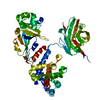
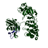
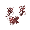
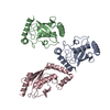
The crystal structure of the protein complex of yeast Hsp40 Sis1 and Hsp70 Ssa1
Components
Heat shock 70 kDa protein cognate 2
SIS1 protein
Keywords
CHAPERONE/PROTEIN TRANSPORT / Hsp40 Sis1 Hsp70 Ssa1 / CHAPERONE-PROTEIN TRANSPORT COMPLEX
Function / homology
Function and homology information
detection of misfolded protein / tRNA import into nucleus / HSP90 chaperone cycle for steroid hormone receptors (SHR) in the presence of ligand / misfolded protein transport / mitochondria-associated ubiquitin-dependent protein catabolic process / nuclear protein quality control by the ubiquitin-proteasome system / misfolded protein binding / response to stress / heat shock protein binding / protein folding chaperone ...detection of misfolded protein / tRNA import into nucleus / HSP90 chaperone cycle for steroid hormone receptors (SHR) in the presence of ligand / misfolded protein transport / mitochondria-associated ubiquitin-dependent protein catabolic process / nuclear protein quality control by the ubiquitin-proteasome system / misfolded protein binding / response to stress / heat shock protein binding / protein folding chaperone / cellular response to starvation / ATP-dependent protein folding chaperone / translational initiation / : / cellular response to heat / protein refolding / protein folding / protein-folding chaperone binding / cytosolic small ribosomal subunit / nucleolus / ATP hydrolysis activity / DNA binding / ATP binding / nucleus / plasma membrane / cytoplasm / cytosol Similarity search - Function
In the structure databanks used in Yorodumi, some data are registered as the other names, "COVID-19 virus" and "2019-nCoV". Here are the details of the virus and the list of structure data.
Jan 31, 2019. EMDB accession codes are about to change! (news from PDBe EMDB page)
EMDB accession codes are about to change! (news from PDBe EMDB page)
The allocation of 4 digits for EMDB accession codes will soon come to an end. Whilst these codes will remain in use, new EMDB accession codes will include an additional digit and will expand incrementally as the available range of codes is exhausted. The current 4-digit format prefixed with “EMD-” (i.e. EMD-XXXX) will advance to a 5-digit format (i.e. EMD-XXXXX), and so on. It is currently estimated that the 4-digit codes will be depleted around Spring 2019, at which point the 5-digit format will come into force.
The EM Navigator/Yorodumi systems omit the EMD- prefix.
Related info.:Q: What is EMD? / ID/Accession-code notation in Yorodumi/EM Navigator
Yorodumi is a browser for structure data from EMDB, PDB, SASBDB, etc.
This page is also the successor to EM Navigator detail page, and also detail information page/front-end page for Omokage search.
The word "yorodu" (or yorozu) is an old Japanese word meaning "ten thousand". "mi" (miru) is to see.
Related info.:EMDB / PDB / SASBDB / Comparison of 3 databanks / Yorodumi Search / Aug 31, 2016. New EM Navigator & Yorodumi / Yorodumi Papers / Jmol/JSmol / Function and homology information / Changes in new EM Navigator and Yorodumi
 Movie
Movie Controller

 Yorodumi
 Open data
 Basic information
 Components
 Keywords
 Function and homology information

 X-RAY DIFFRACTION /
  Authors
 Citation
 Structure visualization
 Downloads & links
 Other downloads
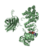


 PDBj
PDBj


 Assembly


 Sample preparation
 / Beamline: 22-ID / Wavelength: 0.9790, 0.9793, 0.9700
 Processing