- PDB-2aya: Solution Structure of the C-Terminal 14 kDa Domain of the tau sub... -
+
Open data
ID or keywords:
Loading...
-
Basic information
Entry
Database: PDB / ID: 2aya
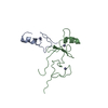
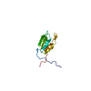
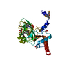
Title
Solution Structure of the C-Terminal 14 kDa Domain of the tau subunit from Escherichia coli DNA Polymerase III
Components
DNA polymerase III subunit tau
Keywords
TRANSFERASE / KH-fold / c-terminus of polymerase III tau subunit
Function / homology
Function and homology information
DNA polymerase III, clamp loader complex / DNA clamp loader activity / DNA polymerase III complex / replisome / DNA polymerase processivity factor activity / DNA-templated DNA replication / ribonucleoside triphosphate phosphatase activity / DNA-directed DNA polymerase / DNA-directed DNA polymerase activity / DNA replication ...DNA polymerase III, clamp loader complex / DNA clamp loader activity / DNA polymerase III complex / replisome / DNA polymerase processivity factor activity / DNA-templated DNA replication / ribonucleoside triphosphate phosphatase activity / DNA-directed DNA polymerase / DNA-directed DNA polymerase activity / DNA replication / viral translational frameshifting / ATP hydrolysis activity / DNA binding / ATP binding / metal ion binding / identical protein binding Similarity search - Function
DNA polymerase III, tau subunit, domain V / DNA polymerase III, tau subunit, domain V / DNA polymerase III subunit tau, DnaB-binding domain IV / DNA polymerase III, tau subunit, domain V superfamily / DNA polymerase III subunits tau domain IV DnaB-binding / DNA polymerase III tau subunit V interacting with alpha / DNA polymerase III, subunit gamma/tau, helical lid domain / DNA polymerase III clamp loader subunit, ATPase lid domain / DNA polymerase III, subunit gamma/ tau, N-terminal / DNA polymerase III, gamma subunit, domain III ...DNA polymerase III, tau subunit, domain V / DNA polymerase III, tau subunit, domain V / DNA polymerase III subunit tau, DnaB-binding domain IV / DNA polymerase III, tau subunit, domain V superfamily / DNA polymerase III subunits tau domain IV DnaB-binding / DNA polymerase III tau subunit V interacting with alpha / DNA polymerase III, subunit gamma/tau, helical lid domain / DNA polymerase III clamp loader subunit, ATPase lid domain / DNA polymerase III, subunit gamma/ tau, N-terminal / DNA polymerase III, gamma subunit, domain III / DNA polymerase III subunits gamma and tau domain III / DNA polymerase III, delta subunit / : / DNA polymerase III, clamp loader complex, gamma/delta/delta subunit, C-terminal / ClpA/B family / GMP Synthetase; Chain A, domain 3 / ATPases associated with a variety of cellular activities / AAA+ ATPase domain / P-loop containing nucleoside triphosphate hydrolase / 2-Layer Sandwich / Alpha Beta Similarity search - Domain/homology
Ionic strength: 100 mM NaCl / pH: 6.8 / Pressure: 1 atm / Temperature: 303 K
-
NMR measurement
Radiation
Protocol: SINGLE WAVELENGTH / Monochromatic (M) / Laue (L): M
Radiation wavelength
Relative weight: 1
NMR spectrometer
Type
Manufacturer
Model
Field strength (MHz)
Spectrometer-ID
Varian INOVA
Varian
INOVA
600
1
Bruker AVANCE
Bruker
AVANCE
800
2
-
Processing
NMR software
Name
Version
Developer
Classification
TopSpin
1.3
Bruker
processing
DYANA
1.5
Guentert
structuresolution
PROSA
6.1
Guentert
processing
OPAL
2.6
Luginbuehl
refinement
CARA
1.2
Damberger
dataanalysis
Refinement
Method: torsion angle dynamics / Software ordinal: 1 Details: The structures are based on a total of 1891 NOE-derived distance constraints and 241 dihedral angle restraints.
NMR representative
Selection criteria: the structure closest to the average
NMR ensemble
Conformer selection criteria: The submitted conformer models are the 20 structures with the lowest energy violations Conformers calculated total number: 200 / Conformers submitted total number: 20
+
About Yorodumi
-
News
-
Feb 9, 2022. New format data for meta-information of EMDB entries
New format data for meta-information of EMDB entries
Version 3 of the EMDB header file is now the official format.
The previous official version 1.9 will be removed from the archive.
In the structure databanks used in Yorodumi, some data are registered as the other names, "COVID-19 virus" and "2019-nCoV". Here are the details of the virus and the list of structure data.
Jan 31, 2019. EMDB accession codes are about to change! (news from PDBe EMDB page)
EMDB accession codes are about to change! (news from PDBe EMDB page)
The allocation of 4 digits for EMDB accession codes will soon come to an end. Whilst these codes will remain in use, new EMDB accession codes will include an additional digit and will expand incrementally as the available range of codes is exhausted. The current 4-digit format prefixed with “EMD-” (i.e. EMD-XXXX) will advance to a 5-digit format (i.e. EMD-XXXXX), and so on. It is currently estimated that the 4-digit codes will be depleted around Spring 2019, at which point the 5-digit format will come into force.
The EM Navigator/Yorodumi systems omit the EMD- prefix.
Related info.:Q: What is EMD? / ID/Accession-code notation in Yorodumi/EM Navigator
Yorodumi is a browser for structure data from EMDB, PDB, SASBDB, etc.
This page is also the successor to EM Navigator detail page, and also detail information page/front-end page for Omokage search.
The word "yorodu" (or yorozu) is an old Japanese word meaning "ten thousand". "mi" (miru) is to see.
Related info.:EMDB / PDB / SASBDB / Comparison of 3 databanks / Yorodumi Search / Aug 31, 2016. New EM Navigator & Yorodumi / Yorodumi Papers / Jmol/JSmol / Function and homology information / Changes in new EM Navigator and Yorodumi
 Movie
Movie Controller

 Yorodumi
 Open data
 Basic information
 Components
 Keywords
 Function and homology information
 Authors
 Citation
 Structure visualization
 Downloads & links
 Other downloads


 PDBj
PDBj
 Assembly
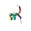
 HNCA
HNCA Sample preparation
 Processing