[English] 日本語
Yorodumi
- PDB-3nf5: Crystal structure of the C-terminal domain of nuclear pore comple... -

+
Open data


ID or keywords:

Loading...

-
Basic information

Entry
Database: PDB / ID: 3nf5
Title
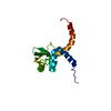
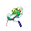
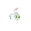
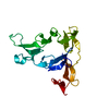
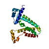
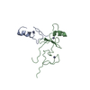
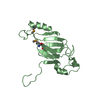
Crystal structure of the C-terminal domain of nuclear pore complex component NUP116 from Candida glabrata
ComponentsNucleoporin NUP116
KeywordsPROTEIN TRANSPORT / Nuclear pore complex / NUP116 / GLEBS domain / STRUCTURAL GENOMICS / PSI-2 / PROTEIN STRUCTURE INITIATIVE / NEW YORK STRUCTURAL GENOMIX RESEARCH CONSORTIUM / NYSGXRC / New York SGX Research Center for Structural Genomics
Function / homology
Function and homology information


structural constituent of nuclear pore / mRNA transport / nuclear pore / protein transport / nuclear membrane
Similarity search - Function
Peptidase S59, nucleoporin / Nuclear pore complex protein NUP98-NUP96 / c-terminal autoproteolytic domain of nucleoporin nup98 / Nucleoporin FG repeat / Nucleoporin FG repeat region / Nuclear pore complex protein Nup98-Nup96-like, autopeptidase S59 domain / Nuclear pore complex protein Nup98-Nup96-like, autopeptidase S59 domain superfamily / Nucleoporin autopeptidase / NUP C-terminal domain profile. / Nucleoporin peptidase S59-like ...Peptidase S59, nucleoporin / Nuclear pore complex protein NUP98-NUP96 / c-terminal autoproteolytic domain of nucleoporin nup98 / Nucleoporin FG repeat / Nucleoporin FG repeat region / Nuclear pore complex protein Nup98-Nup96-like, autopeptidase S59 domain / Nuclear pore complex protein Nup98-Nup96-like, autopeptidase S59 domain superfamily / Nucleoporin autopeptidase / NUP C-terminal domain profile. / Nucleoporin peptidase S59-like / 2-Layer Sandwich / Alpha Beta
Similarity search - Domain/homology
Peptidase S59 domain-containing protein
Similarity search - Component
Biological speciesCandida glabrata (fungus)
MethodX-RAY DIFFRACTION / SYNCHROTRON / MOLECULAR REPLACEMENT / Resolution: 1.94 Å
AuthorsSampathkumar, P. / Manglicmot, D. / Bain, K. / Gilmore, J. / Gheyi, T. / Rout, M. / Sali, A. / Atwell, S. / Thompson, D.A. / Emtage, J.S. ...Sampathkumar, P. / Manglicmot, D. / Bain, K. / Gilmore, J. / Gheyi, T. / Rout, M. / Sali, A. / Atwell, S. / Thompson, D.A. / Emtage, J.S. / Wasserman, S. / Sauder, J.M. / Burley, S.K. / New York SGX Research Center for Structural Genomics (NYSGXRC)
CitationJournal: Proteins / Year: 2012
Title: Atomic structure of the nuclear pore complex targeting domain of a Nup116 homologue from the yeast, Candida glabrata.
Authors: Sampathkumar, P. / Kim, S.J. / Manglicmot, D. / Bain, K.T. / Gilmore, J. / Gheyi, T. / Phillips, J. / Pieper, U. / Fernandez-Martinez, J. / Franke, J.D. / Matsui, T. / Tsuruta, H. / Atwell, ...Authors: Sampathkumar, P. / Kim, S.J. / Manglicmot, D. / Bain, K.T. / Gilmore, J. / Gheyi, T. / Phillips, J. / Pieper, U. / Fernandez-Martinez, J. / Franke, J.D. / Matsui, T. / Tsuruta, H. / Atwell, S. / Thompson, D.A. / Emtage, J.S. / Wasserman, S.R. / Rout, M.P. / Sali, A. / Sauder, J.M. / Almo, S.C. / Burley, S.K.
History
DepositionJun 9, 2010Deposition site: RCSB / Processing site: RCSB
Revision 1.0Aug 4, 2010Provider: repository / Type: Initial release
Revision 1.1Jul 13, 2011Group: Version format compliance
Revision 1.2Dec 21, 2011Group: Structure summary
Revision 1.3Jun 20, 2012Group: Database references
Revision 1.4Oct 10, 2012Group: Database references
Revision 1.5Nov 21, 2018Group: Data collection / Structure summary / Category: audit_author / Item: _audit_author.identifier_ORCID
Revision 1.6Feb 10, 2021Group: Database references / Derived calculations / Structure summary
Category: audit_author / citation_author ...audit_author / citation_author / struct_conn / struct_ref_seq_dif / struct_site
Item: _audit_author.identifier_ORCID / _citation_author.identifier_ORCID ..._audit_author.identifier_ORCID / _citation_author.identifier_ORCID / _struct_conn.pdbx_leaving_atom_flag / _struct_ref_seq_dif.details / _struct_site.pdbx_auth_asym_id / _struct_site.pdbx_auth_comp_id / _struct_site.pdbx_auth_seq_id
Revision 1.7Sep 6, 2023Group: Data collection / Database references / Refinement description
Category: chem_comp_atom / chem_comp_bond ...chem_comp_atom / chem_comp_bond / database_2 / pdbx_initial_refinement_model
Item: _database_2.pdbx_DOI / _database_2.pdbx_database_accession
Revision 1.8Nov 22, 2023Group: Data collection / Category: chem_comp_atom / chem_comp_bond / Item: _chem_comp_atom.atom_id / _chem_comp_bond.atom_id_2

-
Structure visualization

Structure viewerMolecule:
MolmilJmol/JSmol

Downloads & links

-
Assembly

Deposited unit
A: Nucleoporin NUP116
B: Nucleoporin NUP116
hetero molecules


Theoretical massNumber of molelcules
Total (without water)38,0804
Polymers37,8962
Non-polymers1842
Water2,702150
1
A: Nucleoporin NUP116
hetero molecules


Theoretical massNumber of molelcules
Total (without water)19,0402
Polymers18,9481
Non-polymers921
Water181
TypeNameSymmetry operationNumber
identity operation1_555x,y,z1
MethodPISA
2
B: Nucleoporin NUP116
hetero molecules


Theoretical massNumber of molelcules
Total (without water)19,0402
Polymers18,9481
Non-polymers921
Water181
TypeNameSymmetry operationNumber
identity operation1_555x,y,z1
MethodPISA
Unit cell
Length a, b, c (Å)48.703, 67.668, 55.119
Angle α, β, γ (deg.)90.000, 101.370, 90.000
Int Tables number4
Space group name H-MP1211

-
Components

#1: Protein Nucleoporin NUP116 / Strain CBS138 chromosome F complete sequence


Mass: 18947.906 Da / Num. of mol.: 2 / Fragment: C-terminal domain residues 881-1034
Source method: isolated from a genetically manipulated source
Source: (gene. exp.) Candida glabrata (fungus) / Strain: CBS 138 / Gene: CAGL0F06171g, NUP116 (gi50287617) / Plasmid: BC-pSGX3(BC); modified pET26b / Production host: Escherichia coli (E. coli) / Strain (production host): BL21(DE3)-Codon+RIL / References: UniProt: Q6FU56
#2: Chemical ChemComp-GOL / GLYCEROL / GLYCERIN / PROPANE-1,2,3-TRIOL


Mass: 92.094 Da / Num. of mol.: 2 / Source method: obtained synthetically / Formula: C3H8O3
#3: Water ChemComp-HOH / water


Mass: 18.015 Da / Num. of mol.: 150 / Source method: isolated from a natural source / Formula: H2O

-
Experimental details

-
Experiment

ExperimentMethod: X-RAY DIFFRACTION / Number of used crystals: 1

-
Sample preparation

CrystalDensity Matthews: 2.35 Å3/Da / Density % sol: 47.65 %
Crystal growTemperature: 294 K / Method: vapor diffusion, sitting drop / pH: 6.2
Details: 100mM MES, 25% PEGMME 2000, 200mM Sodium potassium tartarae, 0.4microL of 5% ethylacetate used additive into 2microL drop, pH 6.2, VAPOR DIFFUSION, SITTING DROP, temperature 294K

-
Data collection

DiffractionMean temperature: 100 K
Diffraction sourceSource: SYNCHROTRON / Site: APS / Beamline: 31-ID / Wavelength: 0.97929 Å
DetectorType: RAYONIX MX225HE / Detector: CCD / Date: Feb 26, 2010
RadiationMonochromator: diamond / Protocol: SINGLE WAVELENGTH / Monochromatic (M) / Laue (L): M / Scattering type: x-ray
Radiation wavelengthWavelength: 0.97929 Å / Relative weight: 1
ReflectionResolution: 1.94→34.37 Å / Num. obs: 25957 / % possible obs: 99.5 % / Redundancy: 7 % / Biso Wilson estimate: 20 Å2 / Rsym value: 0.14 / Net I/σ(I): 8.3
Reflection shellResolution: 1.94→2.04 Å / Redundancy: 6.9 % / Mean I/σ(I) obs: 3.6 / Rsym value: 0.471 / % possible all: 99.6

-
Processing

Software
NameVersionClassificationNB
REFMAC5.5.0109refinement
PDB_EXTRACT3.1data extraction
MOSFLMdata reduction
SCALAdata scaling
PHASERphasing
RefinementMethod to determine structure: MOLECULAR REPLACEMENT
Starting model: Poly-alanine model of yeast Nup145 PDB ID: 3KEP
Resolution: 1.94→25.09 Å / Cor.coef. Fo:Fc: 0.938 / Cor.coef. Fo:Fc free: 0.91 / Occupancy max: 1 / Occupancy min: 0.5 / SU B: 4.306 / SU ML: 0.124 / Cross valid method: THROUGHOUT / ESU R: 0.173 / ESU R Free: 0.163 / Stereochemistry target values: MAXIMUM LIKELIHOOD / Details: HYDROGENS HAVE BEEN ADDED IN THE RIDING POSITIONS
RfactorNum. reflection% reflectionSelection details
Rfree0.25617 1318 5.1 %RANDOM
Rwork0.20666 ---
obs0.20924 24613 100 %-
Solvent computationIon probe radii: 0.8 Å / Shrinkage radii: 0.8 Å / VDW probe radii: 1.4 Å / Solvent model: BABINET MODEL WITH MASK
Displacement parametersBiso mean: 22.325 Å2
Baniso -1Baniso -2Baniso -3
1-0 Å20 Å20.01 Å2
2---0.01 Å20 Å2
3---0.02 Å2
Refinement stepCycle: LAST / Resolution: 1.94→25.09 Å
ProteinNucleic acidLigandSolventTotal
Num. atoms2394 0 12 150 2556
Refine LS restraints
Refine-IDTypeDev idealDev ideal targetNumber
X-RAY DIFFRACTIONr_bond_refined_d0.020.0222471
X-RAY DIFFRACTIONr_bond_other_d0.0010.021643
X-RAY DIFFRACTIONr_angle_refined_deg1.7811.9863365
X-RAY DIFFRACTIONr_angle_other_deg1.01534041
X-RAY DIFFRACTIONr_dihedral_angle_1_deg6.2755311
X-RAY DIFFRACTIONr_dihedral_angle_2_deg38.46424.571105
X-RAY DIFFRACTIONr_dihedral_angle_3_deg13.31815402
X-RAY DIFFRACTIONr_dihedral_angle_4_deg13.5511510
X-RAY DIFFRACTIONr_chiral_restr0.110.2385
X-RAY DIFFRACTIONr_gen_planes_refined0.0080.0212728
X-RAY DIFFRACTIONr_gen_planes_other0.0010.02470
X-RAY DIFFRACTIONr_nbd_refined
X-RAY DIFFRACTIONr_nbd_other
X-RAY DIFFRACTIONr_nbtor_refined
X-RAY DIFFRACTIONr_nbtor_other
X-RAY DIFFRACTIONr_xyhbond_nbd_refined
X-RAY DIFFRACTIONr_xyhbond_nbd_other
X-RAY DIFFRACTIONr_metal_ion_refined
X-RAY DIFFRACTIONr_metal_ion_other
X-RAY DIFFRACTIONr_symmetry_vdw_refined
X-RAY DIFFRACTIONr_symmetry_vdw_other
X-RAY DIFFRACTIONr_symmetry_hbond_refined
X-RAY DIFFRACTIONr_symmetry_hbond_other
X-RAY DIFFRACTIONr_symmetry_metal_ion_refined
X-RAY DIFFRACTIONr_symmetry_metal_ion_other
X-RAY DIFFRACTIONr_mcbond_it1.1171.51552
X-RAY DIFFRACTIONr_mcbond_other0.3011.5611
X-RAY DIFFRACTIONr_mcangle_it1.99822520
X-RAY DIFFRACTIONr_scbond_it2.9263919
X-RAY DIFFRACTIONr_scangle_it4.5044.5842
X-RAY DIFFRACTIONr_rigid_bond_restr
X-RAY DIFFRACTIONr_sphericity_free
X-RAY DIFFRACTIONr_sphericity_bonded
LS refinement shellResolution: 1.94→1.99 Å / Total num. of bins used: 20
RfactorNum. reflection% reflection
Rfree0.3 95 -
Rwork0.25 1803 -
obs--100 %

+
About Yorodumi

-
News

-
Feb 9, 2022. New format data for meta-information of EMDB entries

New format data for meta-information of EMDB entries

  • Version 3 of the EMDB header file is now the official format.
  • The previous official version 1.9 will be removed from the archive.

Related info.:EMDB header

External links:wwPDB to switch to version 3 of the EMDB data model

-
Aug 12, 2020. Covid-19 info

Covid-19 info

URL: https://pdbjlvh1.pdbj.org/emnavi/covid19.php

New page: Covid-19 featured information page in EM Navigator.

Related info.:Covid-19 info / Mar 5, 2020. Novel coronavirus structure data

+
Mar 5, 2020. Novel coronavirus structure data

Novel coronavirus structure data

Related info.:Yorodumi Speices / Aug 12, 2020. Covid-19 info

External links:COVID-19 featured content - PDBj / Molecule of the Month (242):Coronavirus Proteases

+
Jan 31, 2019. EMDB accession codes are about to change! (news from PDBe EMDB page)

EMDB accession codes are about to change! (news from PDBe EMDB page)

  • The allocation of 4 digits for EMDB accession codes will soon come to an end. Whilst these codes will remain in use, new EMDB accession codes will include an additional digit and will expand incrementally as the available range of codes is exhausted. The current 4-digit format prefixed with “EMD-” (i.e. EMD-XXXX) will advance to a 5-digit format (i.e. EMD-XXXXX), and so on. It is currently estimated that the 4-digit codes will be depleted around Spring 2019, at which point the 5-digit format will come into force.
  • The EM Navigator/Yorodumi systems omit the EMD- prefix.

Related info.:Q: What is EMD? / ID/Accession-code notation in Yorodumi/EM Navigator

External links:EMDB Accession Codes are Changing Soon! / Contact to PDBj

+
Jul 12, 2017. Major update of PDB

Major update of PDB

  • wwPDB released updated PDB data conforming to the new PDBx/mmCIF dictionary.
  • This is a major update changing the version number from 4 to 5, and with Remediation, in which all the entries are updated.
  • In this update, many items about electron microscopy experimental information are reorganized (e.g. em_software).
  • Now, EM Navigator and Yorodumi are based on the updated data.

External links:wwPDB Remediation / Enriched Model Files Conforming to OneDep Data Standards Now Available in the PDB FTP Archive

-
Yorodumi

Thousand views of thousand structures

  • Yorodumi is a browser for structure data from EMDB, PDB, SASBDB, etc.
  • This page is also the successor to EM Navigator detail page, and also detail information page/front-end page for Omokage search.
  • The word "yorodu" (or yorozu) is an old Japanese word meaning "ten thousand". "mi" (miru) is to see.

Related info.:EMDB / PDB / SASBDB / Comparison of 3 databanks / Yorodumi Search / Aug 31, 2016. New EM Navigator & Yorodumi / Yorodumi Papers / Jmol/JSmol / Function and homology information / Changes in new EM Navigator and Yorodumi

Read more