 Movie
Movie Controller

[English] 日本語
 Yorodumi
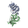
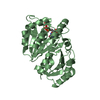
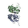
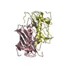
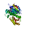
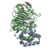
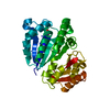
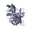
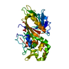
- PDB-2au1: Crystal Structure of group A Streptococcus MAC-1 orthorhombic form -
+ Open data

- Basic information
| Entry | Database: PDB / ID: 2au1 | ||||||
|---|---|---|---|---|---|---|---|
| Title | Crystal Structure of group A Streptococcus MAC-1 orthorhombic form | ||||||
 Components Components | IgG-degrading protease | ||||||
 Keywords Keywords | HYDROLASE / MAC-1 | ||||||
| Function / homology |  Function and homology information Function and homology information | ||||||
| Biological species |  Streptococcus pyogenes (bacteria) Streptococcus pyogenes (bacteria) | ||||||
| Method |  X-RAY DIFFRACTION / SYNCHROTRON / MOLECULAR REPLACEMENT / Resolution: 2.4 Å X-RAY DIFFRACTION / SYNCHROTRON / MOLECULAR REPLACEMENT / Resolution: 2.4 Å | ||||||
 Authors Authors | Agniswamy, J. / Nagiec, M.J. / Liu, M. / Schuck, P. / Musser, J.M. / Sun, P.D. | ||||||
 Citation Citation | Journal: Structure / Year: 2006 Title: Crystal structure of group a streptococcus mac-1: insight into dimer-mediated specificity for recognition of human IgG. Authors: Agniswamy, J. / Nagiec, M.J. / Liu, M. / Schuck, P. / Musser, J.M. / Sun, P.D. | ||||||
| History |
|
- Structure visualization
| Structure viewer | Molecule: MolmilJmol/JSmol |
|---|
- Downloads & links
-Download
| PDBx/mmCIF format | 2au1.cif.gz | 71.1 KB | Display | PDBx/mmCIF format |
|---|---|---|---|---|
| PDB format | pdb2au1.ent.gz | 52.8 KB | Display | PDB format |
| PDBx/mmJSON format | 2au1.json.gz | Tree view | PDBx/mmJSON format | |
| Others |  Other downloads Other downloads |
-Validation report
| Arichive directory | https://data.pdbj.org/pub/pdb/validation_reports/au/2au1ftp://data.pdbj.org/pub/pdb/validation_reports/au/2au1 | HTTPS FTP |
|---|
-Related structure data
-Links
 PDBj
PDBj


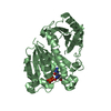
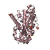
- Assembly
| Deposited unit | 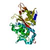
| ||||||||
|---|---|---|---|---|---|---|---|---|---|
| 1 |
| ||||||||
| Unit cell |
|
-Components
| #1: Protein | Mass: 32858.809 Da / Num. of mol.: 1 / Source method: isolated from a natural source / Source: (natural) Streptococcus pyogenes (bacteria) / Strain: MGAS5005 / References: GenBank: 71853263, UniProt: Q7DAM2*PLUS |
|---|---|
| #2: Chemical | ChemComp-BME /   Mass: 78.133 Da / Num. of mol.: 1 / Source method: obtained synthetically / Formula: C2H6OS Mass: 78.133 Da / Num. of mol.: 1 / Source method: obtained synthetically / Formula: C2H6OS |
| #3: Water | ChemComp-HOH /  Mass: 18.015 Da / Num. of mol.: 92 / Source method: isolated from a natural source / Formula: H2O Mass: 18.015 Da / Num. of mol.: 92 / Source method: isolated from a natural source / Formula: H2O |
-Experimental details
-Experiment
| Experiment | Method: X-RAY DIFFRACTION / Number of used crystals: 1 |
|---|
- Sample preparation
| Crystal | Density Matthews: 2.3 Å3/Da / Density % sol: 46 % |
|---|---|
| Crystal grow | Temperature: 277 K / Method: vapor diffusion, hanging drop / pH: 4.8 Details: ammonium sulfate, sodium citrate, pH 4.8, VAPOR DIFFUSION, HANGING DROP, temperature 277K |
-Data collection
| Diffraction | Mean temperature: 100 K |
|---|---|
| Diffraction source | Source: SYNCHROTRON / Site: APS  / Beamline: 22-ID / Wavelength: 0.97954 Å / Beamline: 22-ID / Wavelength: 0.97954 Å |
| Detector | Type: MARMOSAIC 225 mm CCD / Detector: CCD / Date: Nov 7, 2003 |
| Radiation | Protocol: SINGLE WAVELENGTH / Monochromatic (M) / Laue (L): M / Scattering type: x-ray |
| Radiation wavelength | Wavelength: 0.97954 Å / Relative weight: 1 |
| Reflection | Resolution: 2.4→50 Å / Num. all: 13099 / Num. obs: 11324 / % possible obs: 86.4 % / Observed criterion σ(F): 2 / Observed criterion σ(I): 2 / Redundancy: 6.3 % / Rmerge(I) obs: 0.11 |
| Reflection shell | Resolution: 2.4→2.51 Å / % possible all: 5.8 |
- Processing
| Software |
| ||||||||||||||||||||
|---|---|---|---|---|---|---|---|---|---|---|---|---|---|---|---|---|---|---|---|---|---|
| Refinement | Method to determine structure: MOLECULAR REPLACEMENT / Resolution: 2.4→50 Å / σ(F): 0 / Stereochemistry target values: Engh & Huber
| ||||||||||||||||||||
| Refinement step | Cycle: LAST / Resolution: 2.4→50 Å
|
