 Movie
Movie Controller

[English] 日本語
 Yorodumi
- PDB-2arq: Human plasminogen activator inhibitor-2.[loop (66-98) deletion mu... -
+ Open data

- Basic information
| Entry | Database: PDB / ID: 2arq | ||||||
|---|---|---|---|---|---|---|---|
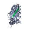
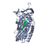
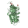
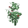
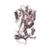
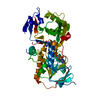
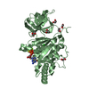
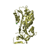
| Title | Human plasminogen activator inhibitor-2.[loop (66-98) deletion mutant] complexed with peptide n-acetyl-teaaagdggvmtgr-oh | ||||||
 Components Components |
| ||||||
 Keywords Keywords | hydrolase inhibitor/peptide / SERPIN / PEPTIDE BINDING / inhibitor / hydrolase inhibitor-peptide complex | ||||||
| Function / homology |  Function and homology information Function and homology informationcornified envelope / Dissolution of Fibrin Clot / fibrinolysis / Gene and protein expression by JAK-STAT signaling after Interleukin-12 stimulation / serine-type endopeptidase inhibitor activity / negative regulation of apoptotic process / : / extracellular region / plasma membrane / cytoplasm Similarity search - Function | ||||||
| Biological species |  Homo sapiens (human) Homo sapiens (human) | ||||||
| Method |  X-RAY DIFFRACTION / MOLECULAR REPLACEMENT / Resolution: 1.85 Å X-RAY DIFFRACTION / MOLECULAR REPLACEMENT / Resolution: 1.85 Å | ||||||
 Authors Authors | Di Giusto, D.A. / Sutherland, A.P. / Jankova, L. / Harrop, S.J. / Curmi, P.M. / King, G.C. | ||||||
 Citation Citation | Journal: J.Mol.Biol. / Year: 2005 Title: Plasminogen activator inhibitor-2 is highly tolerant to P8 residue substitution--implications for serpin mechanistic model and prediction of nsSNP activities Authors: Di Giusto, D.A. / Sutherland, A.P. / Jankova, L. / Harrop, S.J. / Curmi, P.M. / King, G.C. | ||||||
| History |
|
- Structure visualization
| Structure viewer | Molecule: MolmilJmol/JSmol |
|---|
- Downloads & links
-Download
| PDBx/mmCIF format | 2arq.cif.gz | 97.2 KB | Display | PDBx/mmCIF format |
|---|---|---|---|---|
| PDB format | pdb2arq.ent.gz | 72.1 KB | Display | PDB format |
| PDBx/mmJSON format | 2arq.json.gz | Tree view | PDBx/mmJSON format | |
| Others |  Other downloads Other downloads |
-Validation report
| Arichive directory | https://data.pdbj.org/pub/pdb/validation_reports/ar/2arqftp://data.pdbj.org/pub/pdb/validation_reports/ar/2arq | HTTPS FTP |
|---|
-Related structure data
| Related structure data |  2arrC  1jrrS S: Starting model for refinement C: citing same article ( |
|---|---|
| Similar structure data |


-Links
 PDBj
PDBj


- Assembly
| Deposited unit | 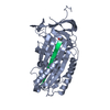
| ||||||||
|---|---|---|---|---|---|---|---|---|---|
| 1 |
| ||||||||
| Unit cell |
|
-Components
| #1: Protein | Mass: 42986.918 Da / Num. of mol.: 1 Source method: isolated from a genetically manipulated source Source: (gene. exp.) Homo sapiens (human) / Production host:  |
|---|---|
| #2: Protein/peptide | Mass: 1319.423 Da / Num. of mol.: 1 / Mutation: T373D Source method: isolated from a genetically manipulated source Source: (gene. exp.) Homo sapiens (human) / Production host: |
| #3: Water | ChemComp-HOH /  Mass: 18.015 Da / Num. of mol.: 345 / Source method: isolated from a natural source / Formula: H2O Mass: 18.015 Da / Num. of mol.: 345 / Source method: isolated from a natural source / Formula: H2O |
| Has protein modification | Y |
-Experimental details
-Experiment
| Experiment | Method: X-RAY DIFFRACTION / Number of used crystals: 1 |
|---|
- Sample preparation
| Crystal | Density Matthews: 2.25 Å3/Da / Density % sol: 45.4 % |
|---|---|
| Crystal grow | Temperature: 293 K / Method: vapor diffusion, sitting drop / pH: 7.5 Details: PEG 8K, pH 7.5, VAPOR DIFFUSION, SITTING DROP, temperature 293K |
-Data collection
| Diffraction | Mean temperature: 100 K |
|---|---|
| Diffraction source | Source: ROTATING ANODE / Type: ENRAF-NONIUS FR571 / Wavelength: 1.5418 Å |
| Detector | Type: MAC Science DIP-2030 / Detector: IMAGE PLATE / Date: Oct 9, 1999 / Details: MIRRORS |
| Radiation | Monochromator: MIRRORS / Protocol: SINGLE WAVELENGTH / Monochromatic (M) / Laue (L): M / Scattering type: x-ray |
| Radiation wavelength | Wavelength: 1.5418 Å / Relative weight: 1 |
| Reflection | Resolution: 1.85→14.8 Å / Num. all: 33539 / Num. obs: 33539 / % possible obs: 96.2 % / Observed criterion σ(F): 0 / Observed criterion σ(I): 0 / Redundancy: 3.9 % / Biso Wilson estimate: 24.5 Å2 / Rmerge(I) obs: 0.069 / Rsym value: 0.069 / Net I/σ(I): 15.2 |
| Reflection shell | Resolution: 1.85→1.92 Å / Redundancy: 3.2 % / Rmerge(I) obs: 0.417 / Mean I/σ(I) obs: 2.1 / Num. unique all: 3330 / Rsym value: 0.417 / % possible all: 98.1 |
- Processing
| Software |
| |||||||||||||||||||||||||||||||||||||||||||||||||||||||||||||||||||||||||||||||||||||||||||||||||||||||||||||||||||||||||||||
|---|---|---|---|---|---|---|---|---|---|---|---|---|---|---|---|---|---|---|---|---|---|---|---|---|---|---|---|---|---|---|---|---|---|---|---|---|---|---|---|---|---|---|---|---|---|---|---|---|---|---|---|---|---|---|---|---|---|---|---|---|---|---|---|---|---|---|---|---|---|---|---|---|---|---|---|---|---|---|---|---|---|---|---|---|---|---|---|---|---|---|---|---|---|---|---|---|---|---|---|---|---|---|---|---|---|---|---|---|---|---|---|---|---|---|---|---|---|---|---|---|---|---|---|---|---|---|
| Refinement | Method to determine structure: MOLECULAR REPLACEMENT Starting model: 1JRR Resolution: 1.85→14.8 Å / Cor.coef. Fo:Fc: 0.959 / Cor.coef. Fo:Fc free: 0.928 / SU B: 3.491 / SU ML: 0.107 / Isotropic thermal model: Isotropic / Cross valid method: THROUGHOUT / σ(F): 0 / σ(I): 0 / ESU R: 0.145 / ESU R Free: 0.151 / Stereochemistry target values: MAXIMUM LIKELIHOOD / Details: HYDROGENS HAVE BEEN ADDED IN THE RIDING POSITIONS
| |||||||||||||||||||||||||||||||||||||||||||||||||||||||||||||||||||||||||||||||||||||||||||||||||||||||||||||||||||||||||||||
| Solvent computation | Ion probe radii: 0.8 Å / Shrinkage radii: 0.8 Å / VDW probe radii: 1.2 Å / Solvent model: MASK | |||||||||||||||||||||||||||||||||||||||||||||||||||||||||||||||||||||||||||||||||||||||||||||||||||||||||||||||||||||||||||||
| Displacement parameters | Biso mean: 22.506 Å2
| |||||||||||||||||||||||||||||||||||||||||||||||||||||||||||||||||||||||||||||||||||||||||||||||||||||||||||||||||||||||||||||
| Refinement step | Cycle: LAST / Resolution: 1.85→14.8 Å
| |||||||||||||||||||||||||||||||||||||||||||||||||||||||||||||||||||||||||||||||||||||||||||||||||||||||||||||||||||||||||||||
| Refine LS restraints |
| |||||||||||||||||||||||||||||||||||||||||||||||||||||||||||||||||||||||||||||||||||||||||||||||||||||||||||||||||||||||||||||
| LS refinement shell | Resolution: 1.852→1.9 Å / Total num. of bins used: 20
|
