- PDB-2ap9: Crystal structure of acetylglutamate kinase from Mycobacterium tu... -
+
Open data
ID or keywords:
Loading...
-
Basic information
Entry
Database: PDB / ID: 2ap9
Title
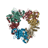
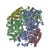
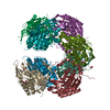
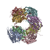
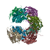
Crystal structure of acetylglutamate kinase from Mycobacterium tuberculosis CDC1551
Components
acetylglutamate kinase
Keywords
SIGNALING PROTEIN / TRANSFERASE / Structural Genomics / Protein Structure Initiative / NYSGXRC / T1702 / acetylglutamate kinase / PSI / New York SGX Research Center for Structural Genomics
Type: ADSC QUANTUM 315 / Detector: CCD / Date: Apr 7, 2005 Details: Vertical focussing mirror down stream of monochromator
Radiation
Monochromator: SAGITALLY FOCUSED Si(111) / Protocol: SINGLE WAVELENGTH / Monochromatic (M) / Laue (L): M / Scattering type: x-ray
Radiation wavelength
Wavelength: 0.975 Å / Relative weight: 1
Reflection
Resolution: 2.8→50 Å / Num. all: 94360 / Num. obs: 94360 / % possible obs: 94 % / Observed criterion σ(I): -1 / Redundancy: 3.17 % / Biso Wilson estimate: 14.5 Å2 / Rsym value: 0.083 / Net I/σ(I): 14.61
Reflection shell
Resolution: 2.8→2.9 Å / Mean I/σ(I) obs: 1.03 / Num. unique all: 6358 / Rsym value: 0.497 / % possible all: 63.5
-
Processing
Software
Name
Version
Classification
DENZO
datareduction
SCALEPACK
datascaling
MLPHARE
phasing
CNS
1.1
refinement
Refinement
Method to determine structure: SAD / Resolution: 2.8→48.8 Å / Rfactor Rfree error: 0.004 / Data cutoff high absF: 178264.75 / Data cutoff low absF: 0 / Isotropic thermal model: RESTRAINED / Cross valid method: THROUGHOUT / σ(F): 0 / Stereochemistry target values: Engh & Huber Details: Certain regions of the model do not have well defined electron density. These are residues 143-152 and 222-231. Two solvent peaks have been refined as Magnesium ions, and one solvent peak ...Details: Certain regions of the model do not have well defined electron density. These are residues 143-152 and 222-231. Two solvent peaks have been refined as Magnesium ions, and one solvent peak has be refined as a Nickle ion (as it is bound to His6 rag).
In the structure databanks used in Yorodumi, some data are registered as the other names, "COVID-19 virus" and "2019-nCoV". Here are the details of the virus and the list of structure data.
Jan 31, 2019. EMDB accession codes are about to change! (news from PDBe EMDB page)
EMDB accession codes are about to change! (news from PDBe EMDB page)
The allocation of 4 digits for EMDB accession codes will soon come to an end. Whilst these codes will remain in use, new EMDB accession codes will include an additional digit and will expand incrementally as the available range of codes is exhausted. The current 4-digit format prefixed with “EMD-” (i.e. EMD-XXXX) will advance to a 5-digit format (i.e. EMD-XXXXX), and so on. It is currently estimated that the 4-digit codes will be depleted around Spring 2019, at which point the 5-digit format will come into force.
The EM Navigator/Yorodumi systems omit the EMD- prefix.
Related info.:Q: What is EMD? / ID/Accession-code notation in Yorodumi/EM Navigator
Yorodumi is a browser for structure data from EMDB, PDB, SASBDB, etc.
This page is also the successor to EM Navigator detail page, and also detail information page/front-end page for Omokage search.
The word "yorodu" (or yorozu) is an old Japanese word meaning "ten thousand". "mi" (miru) is to see.
Related info.:EMDB / PDB / SASBDB / Comparison of 3 databanks / Yorodumi Search / Aug 31, 2016. New EM Navigator & Yorodumi / Yorodumi Papers / Jmol/JSmol / Function and homology information / Changes in new EM Navigator and Yorodumi
 Movie
Movie Controller

 Yorodumi
 Open data
 Basic information
 Components
 Keywords
 Function and homology information
 Mycobacterium tuberculosis (bacteria)
 X-RAY DIFFRACTION /
  Authors
 Citation
 Structure visualization
 Downloads & links
 Other downloads


 PDBj
PDBj Assembly
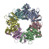

 Mass: 24.305 Da / Num. of mol.: 2 / Source method: obtained synthetically / Formula: Mg
 Mass: 58.693 Da / Num. of mol.: 1 / Source method: obtained synthetically / Formula: Ni
 Mass: 18.015 Da / Num. of mol.: 113 / Source method: isolated from a natural source / Formula: H2O
 Sample preparation
 / Beamline: X29A / Wavelength: 0.975 Å
 Processing