 Movie
Movie Controller


+ Open data

- Basic information
| Entry | Database: PDB / ID: 2anv | ||||||
|---|---|---|---|---|---|---|---|
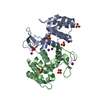
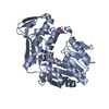
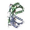
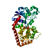
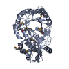
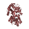
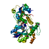
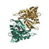
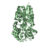
| Title | crystal structure of P22 lysozyme mutant L86M | ||||||
 Components Components | Lysozyme | ||||||
 Keywords Keywords | HYDROLASE / phage lysozyme / direct methods / lanthinide binding sites | ||||||
| Function / homology |  Function and homology information Function and homology informationviral release from host cell by cytolysis / peptidoglycan catabolic process / cell wall macromolecule catabolic process / lysozyme / lysozyme activity / host cell cytoplasm / defense response to bacterium Similarity search - Function | ||||||
| Biological species |  Enterobacteria phage P22 (virus) Enterobacteria phage P22 (virus) | ||||||
| Method |  X-RAY DIFFRACTION / SYNCHROTRON / DIRECT METHODS / Resolution: 1.04 Å X-RAY DIFFRACTION / SYNCHROTRON / DIRECT METHODS / Resolution: 1.04 Å | ||||||
 Authors Authors | Mooers, B.H. / Matthews, B.W. | ||||||
 Citation Citation | Journal: Acta Crystallogr.,Sect.D / Year: 2006 Title: Extension to 2268 atoms of direct methods in the ab initio determination of the unknown structure of bacteriophage P22 lysozyme. Authors: Mooers, B.H. / Matthews, B.W. | ||||||
| History |
|
- Structure visualization
| Structure viewer | Molecule: MolmilJmol/JSmol |
|---|
- Downloads & links
-Download
| PDBx/mmCIF format | 2anv.cif.gz | 157.1 KB | Display | PDBx/mmCIF format |
|---|---|---|---|---|
| PDB format | pdb2anv.ent.gz | 123.4 KB | Display | PDB format |
| PDBx/mmJSON format | 2anv.json.gz | Tree view | PDBx/mmJSON format | |
| Others |  Other downloads Other downloads |
-Validation report
| Arichive directory | https://data.pdbj.org/pub/pdb/validation_reports/an/2anvftp://data.pdbj.org/pub/pdb/validation_reports/an/2anv | HTTPS FTP |
|---|
-Related structure data


-Links
 PDBj
PDBj


- Assembly
| Deposited unit | 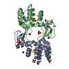
| ||||||||
|---|---|---|---|---|---|---|---|---|---|
| 1 |
| ||||||||
| 2 | 
| ||||||||
| 3 | 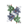
| ||||||||
| Unit cell |
| ||||||||
| Components on special symmetry positions |
|
-Components
-Protein , 1 types, 2 molecules AB
| #1: Protein | Mass: 16178.534 Da / Num. of mol.: 2 / Mutation: L86M Source method: isolated from a genetically manipulated source Source: (gene. exp.) Enterobacteria phage P22 (virus) / Genus: P22-like viruses / Gene: 19 / Production host:  |
|---|
-Non-polymers , 6 types, 500 molecules 


| #2: Chemical | ChemComp-SO4 /  Mass: 96.063 Da / Num. of mol.: 6 / Source method: obtained synthetically / Formula: SO4 Mass: 96.063 Da / Num. of mol.: 6 / Source method: obtained synthetically / Formula: SO4#3: Chemical | ChemComp-MG / |  Mass: 24.305 Da / Num. of mol.: 1 / Source method: obtained synthetically / Formula: Mg Mass: 24.305 Da / Num. of mol.: 1 / Source method: obtained synthetically / Formula: Mg#4: Chemical |  Mass: 150.360 Da / Num. of mol.: 3 / Source method: obtained synthetically / Formula: Sm Mass: 150.360 Da / Num. of mol.: 3 / Source method: obtained synthetically / Formula: Sm#5: Chemical | ChemComp-IOD /  Mass: 126.904 Da / Num. of mol.: 6 / Source method: obtained synthetically / Formula: I Mass: 126.904 Da / Num. of mol.: 6 / Source method: obtained synthetically / Formula: I#6: Chemical |  Mass: 35.453 Da / Num. of mol.: 2 / Source method: obtained synthetically / Formula: Cl Mass: 35.453 Da / Num. of mol.: 2 / Source method: obtained synthetically / Formula: Cl#7: Water | ChemComp-HOH / | Mass: 18.015 Da / Num. of mol.: 482 / Source method: isolated from a natural source / Formula: H2O |
|---|
-Experimental details
-Experiment
| Experiment | Method: X-RAY DIFFRACTION / Number of used crystals: 1 |
|---|
- Sample preparation
| Crystal | Density Matthews: 2.25 Å3/Da / Density % sol: 44.84 % / Description: AB INITIO STRUCTURE DETERMINATION |
|---|---|
| Crystal grow | Temperature: 276 K / pH: 7.5 Details: PEG 3350, pH 7.5, VAPOR DIFFUSION, HANGING DROP, temperature 276K, pH 7.50 |
-Data collection
| Diffraction | Mean temperature: 100 K |
|---|---|
| Diffraction source | Source: SYNCHROTRON / Site: ALS  / Beamline: 8.2.2 / Wavelength: 0.9184 / Beamline: 8.2.2 / Wavelength: 0.9184 |
| Detector | Type: ADSC QUANTUM 315 / Detector: CCD / Date: Oct 10, 2004 / Details: CRYSTAL |
| Radiation | Protocol: SINGLE WAVELENGTH / Monochromatic (M) / Laue (L): M / Scattering type: x-ray |
| Radiation wavelength | Wavelength: 0.9184 Å / Relative weight: 1 |
| Reflection | Resolution: 1.04→42.1 Å / Num. obs: 144532 / % possible obs: 99.6 % / Observed criterion σ(I): -3 / Redundancy: 4.4 % / Biso Wilson estimate: 8.3 Å2 / Rmerge(I) obs: 0.053 / Net I/σ(I): 42.3 |
| Reflection shell | Resolution: 1.04→1.08 Å / Redundancy: 3.1 % / Rmerge(I) obs: 0.222 / Mean I/σ(I) obs: 3.9 / % possible all: 99.4 |
- Processing
| Software |
| |||||||||||||||||||||||||||||||||
|---|---|---|---|---|---|---|---|---|---|---|---|---|---|---|---|---|---|---|---|---|---|---|---|---|---|---|---|---|---|---|---|---|---|---|
| Refinement | Method to determine structure: DIRECT METHODS / Resolution: 1.04→42 Å / σ(F): 0 / Stereochemistry target values: ENGH & HUBER
| |||||||||||||||||||||||||||||||||
| Solvent computation | Solvent model: BABINET | |||||||||||||||||||||||||||||||||
| Refinement step | Cycle: LAST / Resolution: 1.04→42 Å
| |||||||||||||||||||||||||||||||||
| Refine LS restraints |
|
