 Movie
Movie Controller

[English] 日本語
 Yorodumi
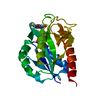
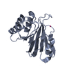
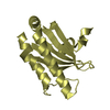
- PDB-2ahd: The Apo structure of Methanococcus jannaschii phosphodiesterase MJ0936 -
+ Open data

- Basic information
| Entry | Database: PDB / ID: 2ahd | ||||||
|---|---|---|---|---|---|---|---|
| Title | The Apo structure of Methanococcus jannaschii phosphodiesterase MJ0936 | ||||||
 Components Components | Phosphodiesterase MJ0936 | ||||||
 Keywords Keywords | HYDROLASE / phosphodiesterase / Structural Genomics / PSI / Protein Structure Initiative / Berkeley Structural Genomics Center / BSGC | ||||||
| Function / homology |  Function and homology information Function and homology informationHydrolases; Acting on ester bonds; Phosphoric-diester hydrolases / hydrolase activity / metal ion binding Similarity search - Function | ||||||
| Biological species |   Methanocaldococcus jannaschii (archaea) Methanocaldococcus jannaschii (archaea) | ||||||
| Method |  X-RAY DIFFRACTION / SYNCHROTRON / MOLECULAR REPLACEMENT / Resolution: 3 Å X-RAY DIFFRACTION / SYNCHROTRON / MOLECULAR REPLACEMENT / Resolution: 3 Å | ||||||
 Authors Authors | Chen, S. / Kim, R. / Kim, S.-H. / Berkeley Structural Genomics Center (BSGC) | ||||||
 Citation Citation | Journal: TO BE PUBLISHED Title: The Apo structure of Methanococcus jannaschii phosphodiesterase MJ0936 Authors: Chen, S. / Kim, R. / Kim, S.-H. | ||||||
| History |
|
- Structure visualization
| Structure viewer | Molecule: MolmilJmol/JSmol |
|---|
- Downloads & links
-Download
| PDBx/mmCIF format | 2ahd.cif.gz | 143 KB | Display | PDBx/mmCIF format |
|---|---|---|---|---|
| PDB format | pdb2ahd.ent.gz | 113.1 KB | Display | PDB format |
| PDBx/mmJSON format | 2ahd.json.gz | Tree view | PDBx/mmJSON format | |
| Others |  Other downloads Other downloads |
-Validation report
| Arichive directory | https://data.pdbj.org/pub/pdb/validation_reports/ah/2ahdftp://data.pdbj.org/pub/pdb/validation_reports/ah/2ahd | HTTPS FTP |
|---|
-Related structure data
| Related structure data |  1s3mS S: Starting model for refinement |
|---|---|
| Similar structure data | |
| Other databases |


-Links
 PDBj
PDBj
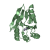
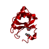
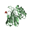
- Assembly
| Deposited unit | 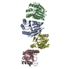
| ||||||||
|---|---|---|---|---|---|---|---|---|---|
| 1 | 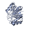
| ||||||||
| 2 | 
| ||||||||
| 3 | 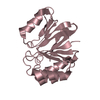
| ||||||||
| 4 | 
| ||||||||
| 5 | 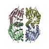
| ||||||||
| Unit cell |
|
-Components
| #1: Protein | Mass: 18931.451 Da / Num. of mol.: 4 Source method: isolated from a genetically manipulated source Source: (gene. exp.) Methanocaldococcus jannaschii (archaea)Plasmid: pSKB3 / Production host:  References: UniProt: Q58346, Hydrolases; Acting on ester bonds; Phosphoric-diester hydrolases #2: Water | ChemComp-HOH / |  Mass: 18.015 Da / Num. of mol.: 149 / Source method: isolated from a natural source / Formula: H2O Mass: 18.015 Da / Num. of mol.: 149 / Source method: isolated from a natural source / Formula: H2O |
|---|
-Experimental details
-Experiment
| Experiment | Method: X-RAY DIFFRACTION / Number of used crystals: 1 |
|---|
- Sample preparation
| Crystal | Density Matthews: 2.7 Å3/Da / Density % sol: 54 % |
|---|---|
| Crystal grow | Temperature: 293 K / Method: vapor diffusion, hanging drop / pH: 8.5 Details: 0.1 M Tris-HCl, pH 8.5, 0.2 M sodium acetate, 30% PEG 4000, VAPOR DIFFUSION, HANGING DROP, temperature 293K |
-Data collection
| Diffraction | Mean temperature: 100 K |
|---|---|
| Diffraction source | Source: SYNCHROTRON / Site: ALS  / Beamline: 5.0.1 / Wavelength: 1 Å / Beamline: 5.0.1 / Wavelength: 1 Å |
| Detector | Type: ADSC QUANTUM 210 / Detector: CCD / Date: Nov 26, 2002 |
| Radiation | Protocol: SINGLE WAVELENGTH / Monochromatic (M) / Laue (L): M / Scattering type: x-ray |
| Radiation wavelength | Wavelength: 1 Å / Relative weight: 1 |
| Reflection | Resolution: 3→50 Å / Num. all: 18648 / Num. obs: 17430 / % possible obs: 93.5 % / Observed criterion σ(F): 0 / Observed criterion σ(I): 0 |
- Processing
| Software |
| ||||||||||||||||||||||||||||||||||||||||||||||||||||||||||||
|---|---|---|---|---|---|---|---|---|---|---|---|---|---|---|---|---|---|---|---|---|---|---|---|---|---|---|---|---|---|---|---|---|---|---|---|---|---|---|---|---|---|---|---|---|---|---|---|---|---|---|---|---|---|---|---|---|---|---|---|---|---|
| Refinement | Method to determine structure: MOLECULAR REPLACEMENT Starting model: pdb entry 1S3M Resolution: 3→44.78 Å / Rfactor Rfree error: 0.007 / Data cutoff high absF: 272853.92 / Data cutoff low absF: 0 / Isotropic thermal model: RESTRAINED / Cross valid method: THROUGHOUT / σ(F): 0
| ||||||||||||||||||||||||||||||||||||||||||||||||||||||||||||
| Solvent computation | Solvent model: FLAT MODEL / Bsol: 54.7722 Å2 / ksol: 0.338479 e/Å3 | ||||||||||||||||||||||||||||||||||||||||||||||||||||||||||||
| Displacement parameters | Biso mean: 78.2 Å2
| ||||||||||||||||||||||||||||||||||||||||||||||||||||||||||||
| Refine analyze |
| ||||||||||||||||||||||||||||||||||||||||||||||||||||||||||||
| Refinement step | Cycle: LAST / Resolution: 3→44.78 Å
| ||||||||||||||||||||||||||||||||||||||||||||||||||||||||||||
| Refine LS restraints |
| ||||||||||||||||||||||||||||||||||||||||||||||||||||||||||||
| LS refinement shell | Resolution: 3→3.19 Å / Rfactor Rfree error: 0.03 / Total num. of bins used: 6
| ||||||||||||||||||||||||||||||||||||||||||||||||||||||||||||
| Xplor file |
|
