 Movie
Movie Controller

[English] 日本語
 Yorodumi
- PDB-2afa: Crystal Structure of putative NAG isomerase from Salmonella typhi... -
+ Open data

- Basic information
| Entry | Database: PDB / ID: 2afa | ||||||
|---|---|---|---|---|---|---|---|
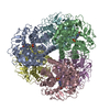
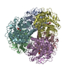
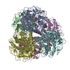
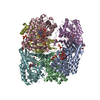
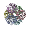
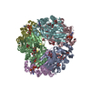
| Title | Crystal Structure of putative NAG isomerase from Salmonella typhimurium | ||||||
 Components Components | NAG isomerase | ||||||
 Keywords Keywords | ISOMERASE / NAG isomerase / Dimer-of-Trimers / renin-binding protein / alpha/alpha-fold / T1489 / Structural Genomics / PSI / Protein Structure Initiative / New York SGX Research Center for Structural Genomics / NYSGXRC | ||||||
| Function / homology |  Function and homology information Function and homology informationsulfoquinovose isomerase / sulfoquinovose isomerase activity / : / carbohydrate metabolic process Similarity search - Function | ||||||
| Biological species |  Salmonella typhimurium (bacteria) Salmonella typhimurium (bacteria) | ||||||
| Method |  X-RAY DIFFRACTION / SYNCHROTRON / SAD / Resolution: 2.15 Å X-RAY DIFFRACTION / SYNCHROTRON / SAD / Resolution: 2.15 Å | ||||||
 Authors Authors | Kumaran, D. / Swaminathan, S. / Burley, S.K. / New York SGX Research Center for Structural Genomics (NYSGXRC) | ||||||
 Citation Citation | Journal: To be Published Title: Crystal Structure of putative NAG isomerase from Salmonella typhimurium Authors: Kumaran, D. / Swaminathan, S. | ||||||
| History |
|
- Structure visualization
| Structure viewer | Molecule: MolmilJmol/JSmol |
|---|
- Downloads & links
-Download
| PDBx/mmCIF format | 2afa.cif.gz | 496 KB | Display | PDBx/mmCIF format |
|---|---|---|---|---|
| PDB format | pdb2afa.ent.gz | 412.2 KB | Display | PDB format |
| PDBx/mmJSON format | 2afa.json.gz | Tree view | PDBx/mmJSON format | |
| Others |  Other downloads Other downloads |
-Validation report
| Arichive directory | https://data.pdbj.org/pub/pdb/validation_reports/af/2afaftp://data.pdbj.org/pub/pdb/validation_reports/af/2afa | HTTPS FTP |
|---|
-Related structure data
| Similar structure data | |
|---|---|
| Other databases |
-Links
 PDBj
PDBj
- Assembly
| Deposited unit | 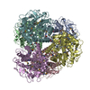
| ||||||||
|---|---|---|---|---|---|---|---|---|---|
| 1 |
| ||||||||
| Unit cell |
|
-Components
| #1: Protein | Mass: 49413.785 Da / Num. of mol.: 6 Source method: isolated from a genetically manipulated source Source: (gene. exp.) Salmonella typhimurium (bacteria) / Gene: yihS / Production host: #2: Water | ChemComp-HOH / |  Mass: 18.015 Da / Num. of mol.: 661 / Source method: isolated from a natural source / Formula: H2O Mass: 18.015 Da / Num. of mol.: 661 / Source method: isolated from a natural source / Formula: H2OHas protein modification | Y | |
|---|
-Experimental details
-Experiment
| Experiment | Method: X-RAY DIFFRACTION / Number of used crystals: 2 |
|---|
- Sample preparation
| Crystal | Density Matthews: 2.2 Å3/Da / Density % sol: 43 % |
|---|---|
| Crystal grow | Temperature: 273 K / Method: vapor diffusion, sitting drop / pH: 6 Details: PEG, sodium cacodylate, sodium acetate , pH 6.0, VAPOR DIFFUSION, SITTING DROP, temperature 273K |
-Data collection
| Diffraction | Mean temperature: 100 K |
|---|---|
| Diffraction source | Source: SYNCHROTRON / Site: NSLS  / Beamline: X25 / Wavelength: 0.979 Å / Beamline: X25 / Wavelength: 0.979 Å |
| Detector | Type: ADSC QUANTUM 315 / Detector: CCD / Date: Oct 19, 2004 / Details: mirrors |
| Radiation | Monochromator: Si 111 CHANNEL / Protocol: SINGLE WAVELENGTH / Monochromatic (M) / Laue (L): M / Scattering type: x-ray |
| Radiation wavelength | Wavelength: 0.979 Å / Relative weight: 1 |
| Reflection | Resolution: 2.15→50 Å / Num. all: 132272 / Num. obs: 132272 / % possible obs: 100 % / Observed criterion σ(F): 0 / Redundancy: 7.4 % / Biso Wilson estimate: 22.5 Å2 / Rmerge(I) obs: 0.08 / Net I/σ(I): 12 |
| Reflection shell | Resolution: 2.15→2.23 Å / Redundancy: 6.8 % / Rmerge(I) obs: 0.75 / Num. unique all: 13231 / % possible all: 100 |
- Processing
| Software |
| |||||||||||||||||||||||||
|---|---|---|---|---|---|---|---|---|---|---|---|---|---|---|---|---|---|---|---|---|---|---|---|---|---|---|
| Refinement | Method to determine structure: SAD / Resolution: 2.15→45.77 Å / Rfactor Rfree error: 0.005 / Data cutoff high absF: 394169.36 / Data cutoff low absF: 0 / Isotropic thermal model: RESTRAINED / Cross valid method: THROUGHOUT / σ(F): 0 / Stereochemistry target values: Engh & Huber Details: In all the chains, residue 416 and residues 376 to 379 were not modelled due to lack of electron density.
| |||||||||||||||||||||||||
| Solvent computation | Solvent model: FLAT MODEL / Bsol: 36.3409 Å2 / ksol: 0.334343 e/Å3 | |||||||||||||||||||||||||
| Displacement parameters | Biso mean: 34 Å2
| |||||||||||||||||||||||||
| Refine analyze |
| |||||||||||||||||||||||||
| Refinement step | Cycle: LAST / Resolution: 2.15→45.77 Å
| |||||||||||||||||||||||||
| Refine LS restraints |
| |||||||||||||||||||||||||
| LS refinement shell | Resolution: 2.15→2.28 Å / Rfactor Rfree error: 0.017 / Total num. of bins used: 6
| |||||||||||||||||||||||||
| Xplor file |
|
