 Movie
Movie Controller

[English] 日本語
 Yorodumi
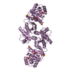
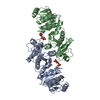
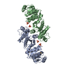
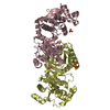
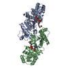
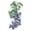
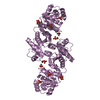
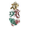
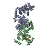
- PDB-2af4: Phosphotransacetylase from Methanosarcina thermophila co-crystall... -
+ Open data

- Basic information
| Entry | Database: PDB / ID: 2af4 | ||||||
|---|---|---|---|---|---|---|---|
| Title | Phosphotransacetylase from Methanosarcina thermophila co-crystallized with coenzyme A | ||||||
 Components Components | Phosphate acetyltransferase | ||||||
 Keywords Keywords | TRANSFERASE / Pta dimer with one CoA ligand bound per monomer / acyltransferase | ||||||
| Function / homology |  Function and homology information Function and homology informationphosphate acetyltransferase / phosphate acetyltransferase activity / acetyl-CoA biosynthetic process / plasma membrane Similarity search - Function | ||||||
| Biological species |   Methanosarcina thermophila (archaea) Methanosarcina thermophila (archaea) | ||||||
| Method |  X-RAY DIFFRACTION / SYNCHROTRON / MOLECULAR REPLACEMENT / Resolution: 2.147 Å X-RAY DIFFRACTION / SYNCHROTRON / MOLECULAR REPLACEMENT / Resolution: 2.147 Å | ||||||
 Authors Authors | Lawrence, S.H. / Luther, K.B. / Ferry, J.G. / Schindelin, H. | ||||||
 Citation Citation | Journal: J.Bacteriol. / Year: 2006 Title: Structural and functional studies suggest a catalytic mechanism for the phosphotransacetylase from Methanosarcina thermophila. Authors: Lawrence, S.H. / Luther, K.B. / Schindelin, H. / Ferry, J.G. #1: Journal: Structure / Year: 2004Title: Crystal structure of phosphotransacetylase from the methanogenic archaeon Methanosarcina thermophila Authors: Iyer, P.P. / Lawrence, S.H. / Luther, K.B. / Rajashankar, K.R. / Yennawar, H.P. / Ferry, J.G. / Schindelin, H. | ||||||
| History |
|
- Structure visualization
| Structure viewer | Molecule: MolmilJmol/JSmol |
|---|
- Downloads & links
-Download
| PDBx/mmCIF format | 2af4.cif.gz | 270.3 KB | Display | PDBx/mmCIF format |
|---|---|---|---|---|
| PDB format | pdb2af4.ent.gz | 222.6 KB | Display | PDB format |
| PDBx/mmJSON format | 2af4.json.gz | Tree view | PDBx/mmJSON format | |
| Others |  Other downloads Other downloads |
-Validation report
| Arichive directory | https://data.pdbj.org/pub/pdb/validation_reports/af/2af4ftp://data.pdbj.org/pub/pdb/validation_reports/af/2af4 | HTTPS FTP |
|---|
-Related structure data
| Related structure data |  2af3C 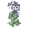 1qztS S: Starting model for refinement C: citing same article ( |
|---|---|
| Similar structure data |
-Links
 PDBj
PDBj


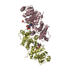
- Assembly
| Deposited unit | 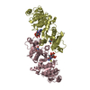
| ||||||||
|---|---|---|---|---|---|---|---|---|---|
| 1 |
| ||||||||
| Unit cell |
|
-Components
| #1: Protein | Mass: 35273.492 Da / Num. of mol.: 2 Source method: isolated from a genetically manipulated source Source: (gene. exp.) Methanosarcina thermophila (archaea) / Gene: pta / Plasmid: pT7-7 / Species (production host): Escherichia coli / Production host:  #2: Chemical |   Mass: 767.534 Da / Num. of mol.: 2 / Source method: obtained synthetically / Formula: C21H36N7O16P3S Mass: 767.534 Da / Num. of mol.: 2 / Source method: obtained synthetically / Formula: C21H36N7O16P3S#3: Water | ChemComp-HOH / |  Mass: 18.015 Da / Num. of mol.: 272 / Source method: isolated from a natural source / Formula: H2O Mass: 18.015 Da / Num. of mol.: 272 / Source method: isolated from a natural source / Formula: H2OHas protein modification | Y | |
|---|
-Experimental details
-Experiment
| Experiment | Method: X-RAY DIFFRACTION / Number of used crystals: 1 |
|---|
- Sample preparation
| Crystal | Density Matthews: 3.08 Å3/Da / Density % sol: 59.75 % |
|---|---|
| Crystal grow | Temperature: 291 K / Method: vapor diffusion, hanging drop / pH: 7.5 Details: sodium citrate, HEPES, Coenzyme A, pH 7.5, VAPOR DIFFUSION, HANGING DROP, temperature 291K |
-Data collection
| Diffraction | Mean temperature: 100 K |
|---|---|
| Diffraction source | Source: SYNCHROTRON / Site: NSLS  / Beamline: X26C / Wavelength: 1.1 Å / Beamline: X26C / Wavelength: 1.1 Å |
| Detector | Type: ADSC QUANTUM 4 / Detector: CCD / Date: Aug 29, 2004 / Details: doubly focusing toroidal mirror |
| Radiation | Monochromator: Si 111 channel / Protocol: SINGLE WAVELENGTH / Monochromatic (M) / Laue (L): M / Scattering type: x-ray |
| Radiation wavelength | Wavelength: 1.1 Å / Relative weight: 1 |
| Reflection | Resolution: 2.147→20 Å / Num. all: 46290 / Num. obs: 45391 / Observed criterion σ(F): 0 / Observed criterion σ(I): 0 / Redundancy: 4.2 % / Rmerge(I) obs: 0.066 / Rsym value: 0.066 / Net I/σ(I): 24 |
| Reflection shell | Resolution: 2.147→2.19 Å / Redundancy: 4.2 % / Rmerge(I) obs: 0.427 / Mean I/σ(I) obs: 2.38 / Rsym value: 0.427 / % possible all: 99.2 |
- Processing
| Software |
| |||||||||||||||||||||||||||||||||||||||||||||||||||||||||||||||||||||||||||||||||||||||||||||||||||||||||||||||||||||||||||||||||||||||||||||||||||||||||||||||||||||||||||||||
|---|---|---|---|---|---|---|---|---|---|---|---|---|---|---|---|---|---|---|---|---|---|---|---|---|---|---|---|---|---|---|---|---|---|---|---|---|---|---|---|---|---|---|---|---|---|---|---|---|---|---|---|---|---|---|---|---|---|---|---|---|---|---|---|---|---|---|---|---|---|---|---|---|---|---|---|---|---|---|---|---|---|---|---|---|---|---|---|---|---|---|---|---|---|---|---|---|---|---|---|---|---|---|---|---|---|---|---|---|---|---|---|---|---|---|---|---|---|---|---|---|---|---|---|---|---|---|---|---|---|---|---|---|---|---|---|---|---|---|---|---|---|---|---|---|---|---|---|---|---|---|---|---|---|---|---|---|---|---|---|---|---|---|---|---|---|---|---|---|---|---|---|---|---|---|---|---|
| Refinement | Method to determine structure: MOLECULAR REPLACEMENT Starting model: PDB ENTRY 1QZT Resolution: 2.147→20 Å / Cor.coef. Fo:Fc: 0.961 / Cor.coef. Fo:Fc free: 0.923 / SU B: 14.715 / SU ML: 0.196 / Cross valid method: THROUGHOUT / ESU R: 0.22 / ESU R Free: 0.21 / Stereochemistry target values: MAXIMUM LIKELIHOOD / Details: HYDROGENS HAVE BEEN ADDED IN THE RIDING POSITIONS
| |||||||||||||||||||||||||||||||||||||||||||||||||||||||||||||||||||||||||||||||||||||||||||||||||||||||||||||||||||||||||||||||||||||||||||||||||||||||||||||||||||||||||||||||
| Solvent computation | Ion probe radii: 0.8 Å / Shrinkage radii: 0.8 Å / VDW probe radii: 1.2 Å / Solvent model: BABINET MODEL WITH MASK | |||||||||||||||||||||||||||||||||||||||||||||||||||||||||||||||||||||||||||||||||||||||||||||||||||||||||||||||||||||||||||||||||||||||||||||||||||||||||||||||||||||||||||||||
| Displacement parameters | Biso mean: 60.125 Å2
| |||||||||||||||||||||||||||||||||||||||||||||||||||||||||||||||||||||||||||||||||||||||||||||||||||||||||||||||||||||||||||||||||||||||||||||||||||||||||||||||||||||||||||||||
| Refinement step | Cycle: LAST / Resolution: 2.147→20 Å
| |||||||||||||||||||||||||||||||||||||||||||||||||||||||||||||||||||||||||||||||||||||||||||||||||||||||||||||||||||||||||||||||||||||||||||||||||||||||||||||||||||||||||||||||
| Refine LS restraints |
| |||||||||||||||||||||||||||||||||||||||||||||||||||||||||||||||||||||||||||||||||||||||||||||||||||||||||||||||||||||||||||||||||||||||||||||||||||||||||||||||||||||||||||||||
| LS refinement shell | Resolution: 2.147→2.202 Å / Total num. of bins used: 20
| |||||||||||||||||||||||||||||||||||||||||||||||||||||||||||||||||||||||||||||||||||||||||||||||||||||||||||||||||||||||||||||||||||||||||||||||||||||||||||||||||||||||||||||||
| Refinement TLS params. | Method: refined / Refine-ID: X-RAY DIFFRACTION
| |||||||||||||||||||||||||||||||||||||||||||||||||||||||||||||||||||||||||||||||||||||||||||||||||||||||||||||||||||||||||||||||||||||||||||||||||||||||||||||||||||||||||||||||
| Refinement TLS group |
|
