 Movie
Movie Controller

[English] 日本語
 Yorodumi
- PDB-2a9v: Crystal structure of a putative gmp synthase subunit a protein (t... -
+ Open data

- Basic information
| Entry | Database: PDB / ID: 2a9v | ||||||
|---|---|---|---|---|---|---|---|
| Title | Crystal structure of a putative gmp synthase subunit a protein (ta0944m) from thermoplasma acidophilum at 2.45 A resolution | ||||||
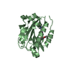
 Components Components | GMP synthase | ||||||
 Keywords Keywords | LIGASE / Structural genomics / Joint Center for Structural Genomics / JCSG / Protein Structure Initiative / PSI-2 | ||||||
| Function / homology |  Function and homology information Function and homology informationGMP synthase activity / GMP synthase (glutamine-hydrolysing) / ATP binding / cytosol Similarity search - Function | ||||||
| Biological species |   Thermoplasma acidophilum (acidophilic) Thermoplasma acidophilum (acidophilic) | ||||||
| Method |  X-RAY DIFFRACTION / SYNCHROTRON / MAD, Molecular replacement / MAD / Resolution: 2.24 Å X-RAY DIFFRACTION / SYNCHROTRON / MAD, Molecular replacement / MAD / Resolution: 2.24 Å | ||||||
 Authors Authors | Joint Center for Structural Genomics (JCSG) | ||||||
 Citation Citation | Journal: To be published Title: Crystal structure of (np_394403.1) from THERMOPLASMA ACIDOPHILUM at 2.45 A resolution Authors: Joint Center for Structural Genomics (JCSG) | ||||||
| History |
|
- Structure visualization
| Structure viewer | Molecule: MolmilJmol/JSmol |
|---|
- Downloads & links
-Download
| PDBx/mmCIF format | 2a9v.cif.gz | 171.2 KB | Display | PDBx/mmCIF format |
|---|---|---|---|---|
| PDB format | pdb2a9v.ent.gz | 134.8 KB | Display | PDB format |
| PDBx/mmJSON format | 2a9v.json.gz | Tree view | PDBx/mmJSON format | |
| Others |  Other downloads Other downloads |
-Validation report
| Arichive directory | https://data.pdbj.org/pub/pdb/validation_reports/a9/2a9vftp://data.pdbj.org/pub/pdb/validation_reports/a9/2a9v | HTTPS FTP |
|---|
-Related structure data
| Related structure data | 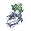 1qdlS S: Starting model for refinement |
|---|---|
| Similar structure data | |
| Other databases |


-Links
 PDBj
PDBj


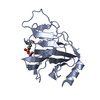
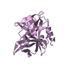
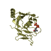
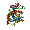
- Assembly
| Deposited unit | 
| ||||||||
|---|---|---|---|---|---|---|---|---|---|
| 1 | 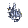
| ||||||||
| 2 | 
| ||||||||
| 3 | 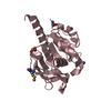
| ||||||||
| 4 | 
| ||||||||
| Unit cell |
|
-Components
| #1: Protein | Mass: 24100.541 Da / Num. of mol.: 4 Source method: isolated from a genetically manipulated source Source: (gene. exp.) Thermoplasma acidophilum (acidophilic) / Gene: guaAA / Production host:  References: UniProt: Q9HJM3, GMP synthase (glutamine-hydrolysing) #2: Chemical | ChemComp-SCN /   Mass: 58.082 Da / Num. of mol.: 5 / Source method: obtained synthetically / Formula: CNS Mass: 58.082 Da / Num. of mol.: 5 / Source method: obtained synthetically / Formula: CNS#3: Chemical | ChemComp-GOL /   Mass: 92.094 Da / Num. of mol.: 7 / Source method: obtained synthetically / Formula: C3H8O3 Mass: 92.094 Da / Num. of mol.: 7 / Source method: obtained synthetically / Formula: C3H8O3#4: Chemical | ChemComp-CL / |   Mass: 35.453 Da / Num. of mol.: 1 / Source method: obtained synthetically / Formula: Cl Mass: 35.453 Da / Num. of mol.: 1 / Source method: obtained synthetically / Formula: Cl#5: Water | ChemComp-HOH / |  Mass: 18.015 Da / Num. of mol.: 211 / Source method: isolated from a natural source / Formula: H2O Mass: 18.015 Da / Num. of mol.: 211 / Source method: isolated from a natural source / Formula: H2OHas protein modification | Y | |
|---|
-Experimental details
-Experiment
| Experiment | Method: X-RAY DIFFRACTION / Number of used crystals: 1 |
|---|
- Sample preparation
| Crystal | Density Matthews: 2 Å3/Da / Density % sol: 37.99 % Description: THE SELENIUM SUBSTRUCTURE WAS SOLVED INITIALLY FROM AN ANOMALOUS DIFFERENCE FOURIER BASED ON A PREVIOUS MOLECULAR REPLACEMENT SOLUTION. THE PHASES WERE THEN REFINED IN SHARP. THE BEST ...Description: THE SELENIUM SUBSTRUCTURE WAS SOLVED INITIALLY FROM AN ANOMALOUS DIFFERENCE FOURIER BASED ON A PREVIOUS MOLECULAR REPLACEMENT SOLUTION. THE PHASES WERE THEN REFINED IN SHARP. THE BEST WARPNTRACE RESULT WAS GENERATED FROM THE MOLECULAR REPLACEMENT MODEL, USING THE MAD PHASES AS PHASE RESTRAINTS FOR REFMAC WITH THE MLHL TARGET.THE SUBSTRUCTURE CAN BE DETERMINED DE NOVO USING SHELXD AND AUTOSHARP. HOWEVER, THE TRACING RESULT IS NOT AS GOOD AS THE TRACE OBTAINED WITH BOTH MAD AND MOLECULAR REPLACEMENT CONTRIBUTIONS. |
|---|---|
| Crystal grow | Temperature: 293 K / Method: vapor diffusion, sitting drop, nanodrop Details: 22.00% PEG 3350, 0.20M NP_Sodium Thiocyanate, VAPOR DIFFUSION,SITTING DROP,NANODROP, temperature 293K |
-Data collection
| Diffraction | Mean temperature: 100 K | |||||||||||||||||||||||||||||||||||||||||||||||||||||||||||||||||||||||||||||||||||||||||||||||||||||||||||||||||||||||||||||||||||||||||||||||||||
|---|---|---|---|---|---|---|---|---|---|---|---|---|---|---|---|---|---|---|---|---|---|---|---|---|---|---|---|---|---|---|---|---|---|---|---|---|---|---|---|---|---|---|---|---|---|---|---|---|---|---|---|---|---|---|---|---|---|---|---|---|---|---|---|---|---|---|---|---|---|---|---|---|---|---|---|---|---|---|---|---|---|---|---|---|---|---|---|---|---|---|---|---|---|---|---|---|---|---|---|---|---|---|---|---|---|---|---|---|---|---|---|---|---|---|---|---|---|---|---|---|---|---|---|---|---|---|---|---|---|---|---|---|---|---|---|---|---|---|---|---|---|---|---|---|---|---|---|---|
| Diffraction source | Source: SYNCHROTRON / Site: ALS  / Beamline: 8.2.2 / Wavelength: 1.00, 0.97971, 0.97947 / Beamline: 8.2.2 / Wavelength: 1.00, 0.97971, 0.97947 | |||||||||||||||||||||||||||||||||||||||||||||||||||||||||||||||||||||||||||||||||||||||||||||||||||||||||||||||||||||||||||||||||||||||||||||||||||
| Detector | Type: ADSC QUANTUM 315 / Detector: CCD / Date: Feb 25, 2005 | |||||||||||||||||||||||||||||||||||||||||||||||||||||||||||||||||||||||||||||||||||||||||||||||||||||||||||||||||||||||||||||||||||||||||||||||||||
| Radiation | Protocol: MAD / Monochromatic (M) / Laue (L): M / Scattering type: x-ray | |||||||||||||||||||||||||||||||||||||||||||||||||||||||||||||||||||||||||||||||||||||||||||||||||||||||||||||||||||||||||||||||||||||||||||||||||||
| Radiation wavelength |
| |||||||||||||||||||||||||||||||||||||||||||||||||||||||||||||||||||||||||||||||||||||||||||||||||||||||||||||||||||||||||||||||||||||||||||||||||||
| Reflection | Resolution: 2.24→66.27 Å / Num. obs: 29096 / % possible obs: 78.6 % / Redundancy: 3.2 % / Rmerge(I) obs: 0.053 / Rsym value: 0.053 / Net I/σ(I): 9 | |||||||||||||||||||||||||||||||||||||||||||||||||||||||||||||||||||||||||||||||||||||||||||||||||||||||||||||||||||||||||||||||||||||||||||||||||||
| Reflection shell | Diffraction-ID: 1
|
-Phasing
| Phasing | Method: MAD |
|---|
- Processing
| Software |
| |||||||||||||||||||||||||||||||||||||||||||||||||||||||||||||||||||||||||||||||||||||||||||||||||||||||||||||||||||||||||||||
|---|---|---|---|---|---|---|---|---|---|---|---|---|---|---|---|---|---|---|---|---|---|---|---|---|---|---|---|---|---|---|---|---|---|---|---|---|---|---|---|---|---|---|---|---|---|---|---|---|---|---|---|---|---|---|---|---|---|---|---|---|---|---|---|---|---|---|---|---|---|---|---|---|---|---|---|---|---|---|---|---|---|---|---|---|---|---|---|---|---|---|---|---|---|---|---|---|---|---|---|---|---|---|---|---|---|---|---|---|---|---|---|---|---|---|---|---|---|---|---|---|---|---|---|---|---|---|
| Refinement | Method to determine structure: MAD, Molecular replacement Starting model: 1qdl Resolution: 2.24→66.27 Å / Cor.coef. Fo:Fc: 0.953 / Cor.coef. Fo:Fc free: 0.9 / SU B: 12.757 / SU ML: 0.157 / Cross valid method: THROUGHOUT / ESU R: 0.952 / ESU R Free: 0.295 Stereochemistry target values: MAXIMUM LIKELIHOOD WITH PHASES Details: HYDROGENS HAVE BEEN ADDED IN THE RIDING POSITIONS. THE NOMINAL RESOLUTION IS 2.45 A WITH 3445 OBSERVED REFLECTIONS BETWEEN 2.45-2.24 (40.4% COMPLETE FOR THIS SHELL) INCLUDED IN THE REFINEMENT.
| |||||||||||||||||||||||||||||||||||||||||||||||||||||||||||||||||||||||||||||||||||||||||||||||||||||||||||||||||||||||||||||
| Solvent computation | Ion probe radii: 0.8 Å / Shrinkage radii: 0.8 Å / VDW probe radii: 1.2 Å / Solvent model: MASK | |||||||||||||||||||||||||||||||||||||||||||||||||||||||||||||||||||||||||||||||||||||||||||||||||||||||||||||||||||||||||||||
| Displacement parameters | Biso mean: 25.529 Å2
| |||||||||||||||||||||||||||||||||||||||||||||||||||||||||||||||||||||||||||||||||||||||||||||||||||||||||||||||||||||||||||||
| Refinement step | Cycle: LAST / Resolution: 2.24→66.27 Å
| |||||||||||||||||||||||||||||||||||||||||||||||||||||||||||||||||||||||||||||||||||||||||||||||||||||||||||||||||||||||||||||
| Refine LS restraints |
| |||||||||||||||||||||||||||||||||||||||||||||||||||||||||||||||||||||||||||||||||||||||||||||||||||||||||||||||||||||||||||||
| LS refinement shell | Resolution: 2.24→2.298 Å / Total num. of bins used: 20
|
