 Movie
Movie Controller


+ Open data

- Basic information
| Entry | Database: PDB / ID: 2a8m | ||||||
|---|---|---|---|---|---|---|---|
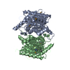
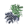
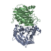
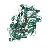
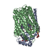
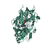
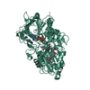
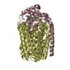
| Title | Crystal Structure of Human Taspase1 (T234S mutant) | ||||||
 Components Components | Threonine aspartase 1 | ||||||
 Keywords Keywords | HYDROLASE / Taspase / MLL / Leukemia / Asparaginase | ||||||
| Function / homology |  Function and homology information Function and homology informationHydrolases; Acting on peptide bonds (peptidases); Threonine endopeptidases / Formation of WDR5-containing histone-modifying complexes / threonine-type endopeptidase activity / protein maturation / positive regulation of DNA-templated transcription / proteolysis / identical protein binding / cytoplasm / cytosol Similarity search - Function | ||||||
| Biological species |  Homo sapiens (human) Homo sapiens (human) | ||||||
| Method |  X-RAY DIFFRACTION / SYNCHROTRON / SAD / Resolution: 2.6 Å X-RAY DIFFRACTION / SYNCHROTRON / SAD / Resolution: 2.6 Å | ||||||
 Authors Authors | Khan, J.A. / Dunn, B.M. / Tong, L. | ||||||
 Citation Citation | Journal: Structure / Year: 2005 Title: Crystal Structure of Human Taspase1, a Crucial Protease Regulating the Function of MLL. Authors: Khan, J.A. / Dunn, B.M. / Tong, L. | ||||||
| History |
|
- Structure visualization
| Structure viewer | Molecule: MolmilJmol/JSmol |
|---|
- Downloads & links
-Download
| PDBx/mmCIF format | 2a8m.cif.gz | 127.1 KB | Display | PDBx/mmCIF format |
|---|---|---|---|---|
| PDB format | pdb2a8m.ent.gz | 97.9 KB | Display | PDB format |
| PDBx/mmJSON format | 2a8m.json.gz | Tree view | PDBx/mmJSON format | |
| Others |  Other downloads Other downloads |
-Validation report
| Arichive directory | https://data.pdbj.org/pub/pdb/validation_reports/a8/2a8mftp://data.pdbj.org/pub/pdb/validation_reports/a8/2a8m | HTTPS FTP |
|---|
-Related structure data


-Links
 PDBj
PDBj


- Assembly
| Deposited unit | 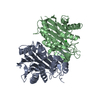
| ||||||||
|---|---|---|---|---|---|---|---|---|---|
| 1 |
| ||||||||
| Unit cell |
|
-Components
| #1: Protein | Mass: 44499.441 Da / Num. of mol.: 2 / Mutation: T234S Source method: isolated from a genetically manipulated source Source: (gene. exp.) Homo sapiens (human) / Gene: C20orf13 / Production host:  References: UniProt: Q9H6P5, Hydrolases; Acting on peptide bonds (peptidases); Threonine endopeptidases #2: Chemical |   Mass: 35.453 Da / Num. of mol.: 2 / Source method: obtained synthetically / Formula: Cl Mass: 35.453 Da / Num. of mol.: 2 / Source method: obtained synthetically / Formula: Cl#3: Water | ChemComp-HOH / |  Mass: 18.015 Da / Num. of mol.: 109 / Source method: isolated from a natural source / Formula: H2O Mass: 18.015 Da / Num. of mol.: 109 / Source method: isolated from a natural source / Formula: H2O |
|---|
-Experimental details
-Experiment
| Experiment | Method: X-RAY DIFFRACTION / Number of used crystals: 1 |
|---|
- Sample preparation
| Crystal | Density Matthews: 1.8 Å3/Da / Density % sol: 30 % |
|---|---|
| Crystal grow | Temperature: 294 K / pH: 6 Details: pH 6.0, VAPOR DIFFUSION, SITTING DROP, temperature 294K |
-Data collection
| Diffraction | Mean temperature: 100 K |
|---|---|
| Diffraction source | Source: SYNCHROTRON / Site: NSLS  / Beamline: X4A / Wavelength: 0.9798 / Beamline: X4A / Wavelength: 0.9798 |
| Detector | Type: ADSC QUANTUM 4 / Detector: CCD / Date: Feb 5, 2005 / Details: MIRRORS |
| Radiation | Monochromator: SI(111) / Protocol: SINGLE WAVELENGTH / Monochromatic (M) / Laue (L): M / Scattering type: x-ray |
| Radiation wavelength | Wavelength: 0.9798 Å / Relative weight: 1 |
| Reflection | Resolution: 2.6→30 Å / Num. obs: 16867 / % possible obs: 93.6 % / Redundancy: 5.9 % / Biso Wilson estimate: 34.6 Å2 / Rmerge(I) obs: 0.101 / Net I/σ(I): 15.0097 |
| Reflection shell | Resolution: 2.6→2.69 Å / Redundancy: 4.8 % / Rmerge(I) obs: 0.392 / Mean I/σ(I) obs: 2.96 / % possible all: 73.1 |
- Processing
| Software |
| ||||||||||||||||||||||||||||||||||||||||||||||||||||||||||||
|---|---|---|---|---|---|---|---|---|---|---|---|---|---|---|---|---|---|---|---|---|---|---|---|---|---|---|---|---|---|---|---|---|---|---|---|---|---|---|---|---|---|---|---|---|---|---|---|---|---|---|---|---|---|---|---|---|---|---|---|---|---|
| Refinement | Method to determine structure: SAD / Resolution: 2.6→29.59 Å / Rfactor Rfree error: 0.007 / Data cutoff high absF: 245503.56 / Data cutoff low absF: 0 / Isotropic thermal model: RESTRAINED / Cross valid method: THROUGHOUT
| ||||||||||||||||||||||||||||||||||||||||||||||||||||||||||||
| Solvent computation | Solvent model: FLAT MODEL / Bsol: 41.0479 Å2 / ksol: 0.349278 e/Å3 | ||||||||||||||||||||||||||||||||||||||||||||||||||||||||||||
| Displacement parameters | Biso mean: 45.9 Å2
| ||||||||||||||||||||||||||||||||||||||||||||||||||||||||||||
| Refine analyze |
| ||||||||||||||||||||||||||||||||||||||||||||||||||||||||||||
| Refinement step | Cycle: LAST / Resolution: 2.6→29.59 Å
| ||||||||||||||||||||||||||||||||||||||||||||||||||||||||||||
| Refine LS restraints |
| ||||||||||||||||||||||||||||||||||||||||||||||||||||||||||||
| Refine LS restraints NCS | NCS model details: CONSTR | ||||||||||||||||||||||||||||||||||||||||||||||||||||||||||||
| LS refinement shell | Resolution: 2.6→2.69 Å / Rfactor Rfree error: 0.034 / Total num. of bins used: 10
| ||||||||||||||||||||||||||||||||||||||||||||||||||||||||||||
| Xplor file |
|
