 Movie
Movie Controller

[English] 日本語
 Yorodumi
- PDB-2a4v: Crystal Structure of a truncated mutant of yeast nuclear thiol pe... -
+ Open data

- Basic information
| Entry | Database: PDB / ID: 2a4v | ||||||
|---|---|---|---|---|---|---|---|
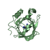
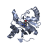
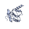
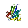
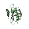
| Title | Crystal Structure of a truncated mutant of yeast nuclear thiol peroxidase | ||||||
 Components Components | Peroxiredoxin DOT5 | ||||||
 Keywords Keywords | OXIDOREDUCTASE / yeast nuclear thiol peroxidase / atypical 2-Cys peroxiredoxin | ||||||
| Function / homology |  Function and homology information Function and homology informationthioredoxin-dependent peroxiredoxin / thioredoxin peroxidase activity / cell redox homeostasis / cellular response to oxidative stress / chromosome, telomeric region / nucleus / cytoplasm Similarity search - Function | ||||||
| Biological species |  | ||||||
| Method |  X-RAY DIFFRACTION / SYNCHROTRON / MOLECULAR REPLACEMENT / Resolution: 1.8 Å X-RAY DIFFRACTION / SYNCHROTRON / MOLECULAR REPLACEMENT / Resolution: 1.8 Å | ||||||
 Authors Authors | Choi, J. / Choi, S. / Chon, J.-K. / Choi, J. / Cha, M.-K. / Kim, I.-H. / Shin, W. | ||||||
 Citation Citation | Journal: Proteins / Year: 2005 Title: Crystal structure of the C107S/C112S mutant of yeast nuclear 2-Cys peroxiredoxin Authors: Choi, J. / Choi, S. / Chon, J.-K. / Choi, J. / Cha, M.-K. / Kim, I.-H. / Shin, W. | ||||||
| History |
|
- Structure visualization
| Structure viewer | Molecule: MolmilJmol/JSmol |
|---|
- Downloads & links
-Download
| PDBx/mmCIF format | 2a4v.cif.gz | 45.9 KB | Display | PDBx/mmCIF format |
|---|---|---|---|---|
| PDB format | pdb2a4v.ent.gz | 32.1 KB | Display | PDB format |
| PDBx/mmJSON format | 2a4v.json.gz | Tree view | PDBx/mmJSON format | |
| Others |  Other downloads Other downloads |
-Validation report
| Arichive directory | https://data.pdbj.org/pub/pdb/validation_reports/a4/2a4vftp://data.pdbj.org/pub/pdb/validation_reports/a4/2a4v | HTTPS FTP |
|---|
-Related structure data
| Similar structure data |
|---|

-Links
 PDBj
PDBj- Assembly
| Deposited unit | 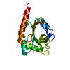
| ||||||||
|---|---|---|---|---|---|---|---|---|---|
| 1 |
| ||||||||
| Unit cell |
|
-Components
| #1: Protein | Mass: 17864.195 Da / Num. of mol.: 1 / Fragment: C-TERMINAL DOMAIN / Mutation: C107S/C112S/K123E Source method: isolated from a genetically manipulated source Source: (gene. exp.) Gene: DOT5 / Plasmid: pT7-7 / Species (production host): Escherichia coli / Production host:  |
|---|---|
| #2: Water | ChemComp-HOH /  Mass: 18.015 Da / Num. of mol.: 156 / Source method: isolated from a natural source / Formula: H2O Mass: 18.015 Da / Num. of mol.: 156 / Source method: isolated from a natural source / Formula: H2O |
-Experimental details
-Experiment
| Experiment | Method: X-RAY DIFFRACTION / Number of used crystals: 1 |
|---|
- Sample preparation
| Crystal | Density Matthews: 1.91 Å3/Da / Density % sol: 35.5 % | |||||||||||||||||||||||||||||||||||
|---|---|---|---|---|---|---|---|---|---|---|---|---|---|---|---|---|---|---|---|---|---|---|---|---|---|---|---|---|---|---|---|---|---|---|---|---|
| Crystal grow | Temperature: 283 K / Method: vapor diffusion, hanging drop / pH: 7.9 Details: PEG 3350, mercury(II) acetate, Tris-HCl, pH 7.9, VAPOR DIFFUSION, HANGING DROP, temperature 283K | |||||||||||||||||||||||||||||||||||
| Crystal grow | *PLUS pH: 7.4 / Details: Choi, J., (2005) Acta Crystallogr., F61, 659. | |||||||||||||||||||||||||||||||||||
| Components of the solutions | *PLUS
|
-Data collection
| Diffraction | Mean temperature: 100 K |
|---|---|
| Diffraction source | Source: SYNCHROTRON / Site: PAL/PLS  / Beamline: 6B / Wavelength: 1.12714 Å / Beamline: 6B / Wavelength: 1.12714 Å |
| Detector | Type: BRUKER PROTEUM 300 / Detector: CCD / Date: Jan 1, 2005 |
| Radiation | Monochromator: double crystal Monochromator / Protocol: SINGLE WAVELENGTH / Monochromatic (M) / Laue (L): M / Scattering type: x-ray |
| Radiation wavelength | Wavelength: 1.12714 Å / Relative weight: 1 |
| Reflection | Resolution: 1.8→20 Å / Num. all: 12169 / Num. obs: 11928 / % possible obs: 98.1 % / Observed criterion σ(F): 1 / Observed criterion σ(I): 1 / Redundancy: 4.6 % / Biso Wilson estimate: 20 Å2 / Rmerge(I) obs: 0.096 / Rsym value: 0.085 / Χ2: 4.97 / Net I/σ(I): 12.4 |
| Reflection shell | Resolution: 1.8→1.86 Å / % possible obs: 97 % / Redundancy: 4.6 % / Rmerge(I) obs: 0.362 / Mean I/σ(I) obs: 3.09 / Num. measured obs: 1173 / Num. unique all: 1171 / Rsym value: 0.321 / Χ2: 2.486 / % possible all: 97 |
| Reflection | *PLUS Highest resolution: 1.8 Å / Lowest resolution: 20 Å / Num. measured all: 55862 |
| Reflection shell | *PLUS % possible obs: 97 % / Num. unique obs: 1171 / Num. measured obs: 5457 / Mean I/σ(I) obs: 3 |
-Phasing
| Phasing MR | Rfactor: 0.57 / Cor.coef. Fo:Fc: 0.153
|
|---|
- Processing
| Software |
| ||||||||||||||||||||||||||||||||||||||||||||||||||||||||||||||||||||||||||||||||||||||||||
|---|---|---|---|---|---|---|---|---|---|---|---|---|---|---|---|---|---|---|---|---|---|---|---|---|---|---|---|---|---|---|---|---|---|---|---|---|---|---|---|---|---|---|---|---|---|---|---|---|---|---|---|---|---|---|---|---|---|---|---|---|---|---|---|---|---|---|---|---|---|---|---|---|---|---|---|---|---|---|---|---|---|---|---|---|---|---|---|---|---|---|---|
| Refinement | Method to determine structure: MOLECULAR REPLACEMENT / Resolution: 1.8→20 Å / Cor.coef. Fo:Fc: 0.963 / Cor.coef. Fo:Fc free: 0.934 / SU B: 2.856 / SU ML: 0.09 / Cross valid method: THROUGHOUT / σ(F): 0 / ESU R: 0.149 / ESU R Free: 0.139 / Stereochemistry target values: MAXIMUM LIKELIHOOD
| ||||||||||||||||||||||||||||||||||||||||||||||||||||||||||||||||||||||||||||||||||||||||||
| Solvent computation | Ion probe radii: 0.8 Å / Shrinkage radii: 0.8 Å / VDW probe radii: 1.2 Å / Solvent model: MASK | ||||||||||||||||||||||||||||||||||||||||||||||||||||||||||||||||||||||||||||||||||||||||||
| Displacement parameters | Biso mean: 19.279 Å2
| ||||||||||||||||||||||||||||||||||||||||||||||||||||||||||||||||||||||||||||||||||||||||||
| Refinement step | Cycle: LAST / Resolution: 1.8→20 Å
| ||||||||||||||||||||||||||||||||||||||||||||||||||||||||||||||||||||||||||||||||||||||||||
| Refine LS restraints |
| ||||||||||||||||||||||||||||||||||||||||||||||||||||||||||||||||||||||||||||||||||||||||||
| LS refinement shell | Resolution: 1.8→1.846 Å / Total num. of bins used: 20
| ||||||||||||||||||||||||||||||||||||||||||||||||||||||||||||||||||||||||||||||||||||||||||
| Software | *PLUS Name: REFMAC / Version: 5.2.0005 / Classification: refinement | ||||||||||||||||||||||||||||||||||||||||||||||||||||||||||||||||||||||||||||||||||||||||||
| Refinement | *PLUS Highest resolution: 1.8 Å / Lowest resolution: 20 Å / % reflection Rfree: 5 % / Rfactor Rwork: 0.167 | ||||||||||||||||||||||||||||||||||||||||||||||||||||||||||||||||||||||||||||||||||||||||||
| Solvent computation | *PLUS | ||||||||||||||||||||||||||||||||||||||||||||||||||||||||||||||||||||||||||||||||||||||||||
| Displacement parameters | *PLUS Biso mean: 18 Å2 | ||||||||||||||||||||||||||||||||||||||||||||||||||||||||||||||||||||||||||||||||||||||||||
| Refine LS restraints | *PLUS
|
